2F9W image
Deposition Date 2005-12-06
Release Date 2006-08-22
Last Version Date 2024-10-30
Entry Detail
PDB ID:
2F9W
Keywords:
Title:
Structure of the type III CoaA from Pseudomonas aeruginosa
Biological Source:
Source Organism:
Host Organism:
Method Details:
Experimental Method:
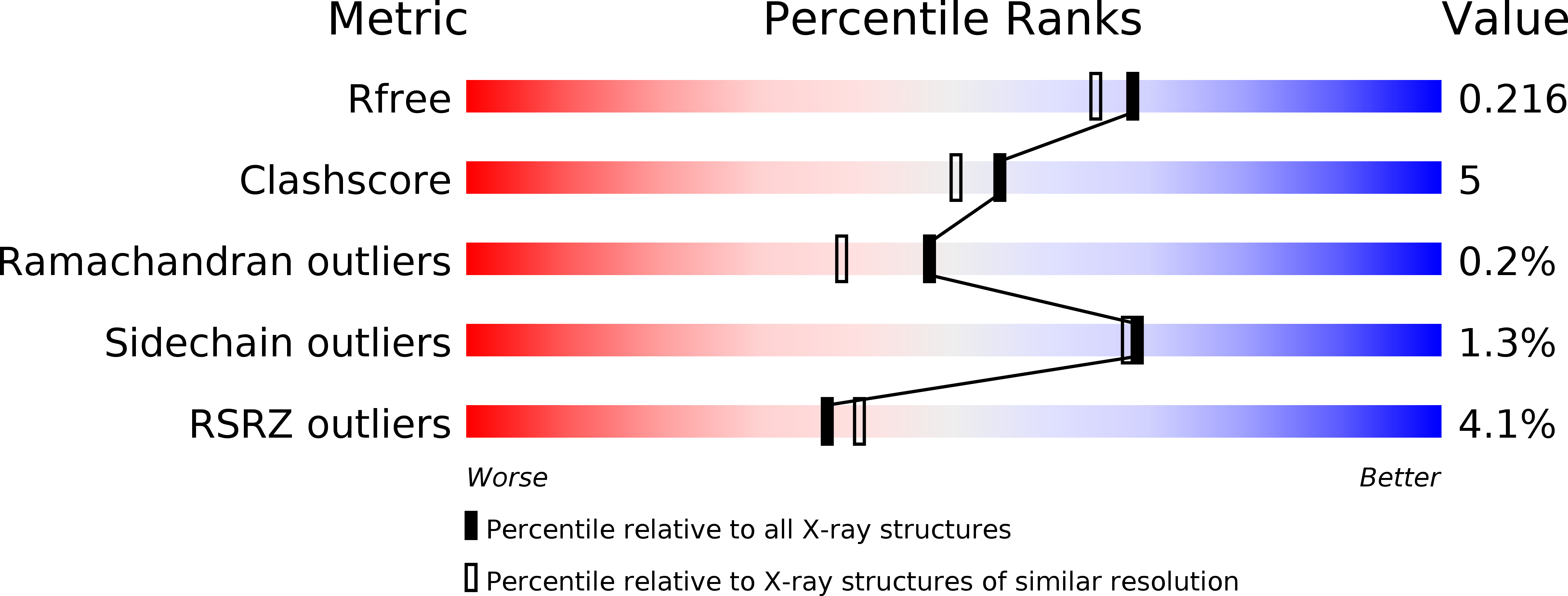
Resolution:
1.90 Å
R-Value Free:
0.22
R-Value Work:
0.21
R-Value Observed:
0.21
Space Group:
P 41 21 2
Macromolecular Entities
Polymer Type:polypeptide(L)
Molecule:Pantothenate Kinase
Gene (Uniprot):coaX
Chain IDs:A, B
Chain Length:271
Number of Molecules:2
Biological Source:Pseudomonas aeruginosa
Modified Residue
Compound ID Chain ID Parent Comp ID Details 2D Image
MSE A MET SELENOMETHIONINE
Primary Citation
Prokaryotic Type II and Type III Pantothenate Kinases: The Same Monomer Fold Creates Dimers with Distinct Catalytic Properties.
Structure 14 1251 1261 (2006)
PMID: 16905099 DOI: 10.1016/j.str.2006.06.008

Abstact

Three distinct isoforms of pantothenate kinase (CoaA) in bacteria catalyze the first step in coenzyme A biosynthesis. The structures of the type II (Staphylococcus aureus, SaCoaA) and type III (Pseudomonas aeruginosa, PaCoaA) enzymes reveal that they assemble nearly identical subunits with actin-like folds into dimers that exhibit distinct biochemical properties. PaCoaA has a fully enclosed pantothenate binding pocket and requires a monovalent cation to weakly bind ATP in an open cavity that does not interact with the adenine nucleotide. Pantothenate binds to an open pocket in SaCoaA that strongly binds ATP by using a classical P loop architecture coupled with specific interactions with the adenine moiety. The PaCoaA*Pan binary complex explains the resistance of bacteria possessing this isoform to the pantothenamide antibiotics, and the similarity between SaCoaA and human pantothenate kinase 2 explains the molecular basis for the development of the neurodegenerative phenotype in three mutations in the human protein.

Legend

Protein

Chemical

Disease

Primary Citation of related structures