2F91 image
Deposition Date 2005-12-05
Release Date 2006-04-18
Last Version Date 2024-11-13
Entry Detail
PDB ID:
2F91
Title:
1.2A resolution structure of a crayfish trypsin complexed with a peptide inhibitor, SGTI
Biological Source:
Source Organism:
(Taxon ID: )Pontastacus leptodactylus (Taxon ID: 6717)
Method Details:
Experimental Method:
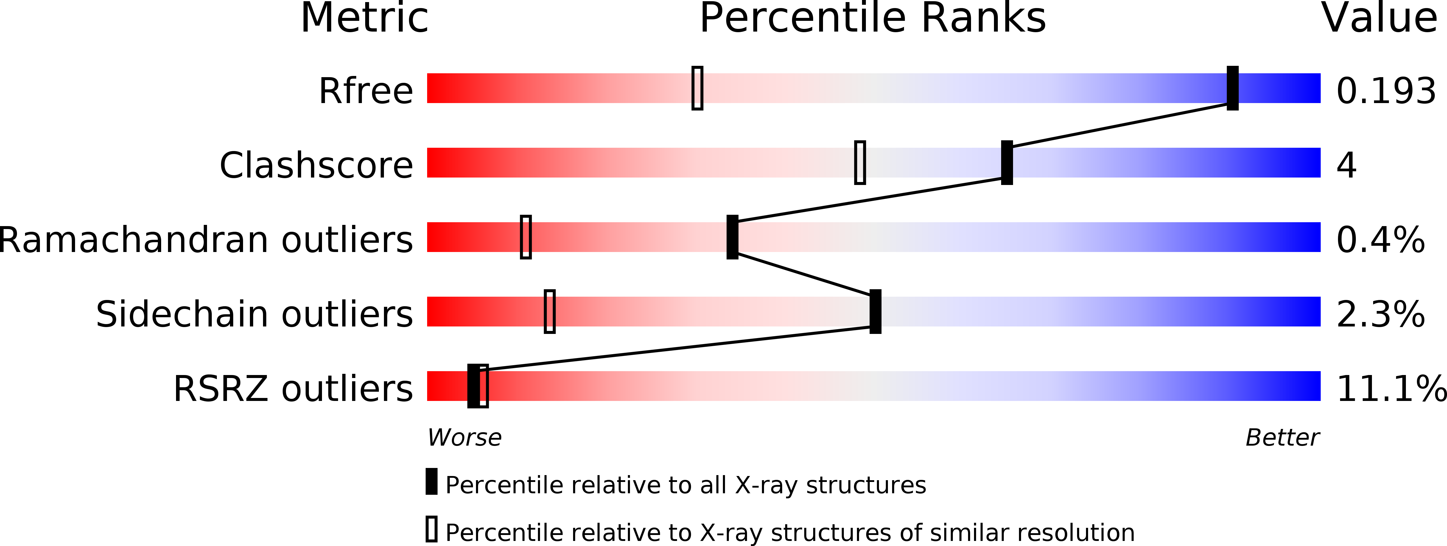
Resolution:
1.20 Å
R-Value Free:
0.18
Space Group:
P 21 21 21
Macromolecular Entities
Polymer Type:polypeptide(L)
Molecule:hepatopancreas trypsin
Chain IDs:A
Chain Length:237
Number of Molecules:1
Biological Source:Pontastacus leptodactylus
Polymer Type:polypeptide(L)
Molecule:Serine protease inhibitor I/II
Chain IDs:B
Chain Length:35
Number of Molecules:1
Biological Source:
Primary Citation
Enzyme:Substrate Hydrogen Bond Shortening during the Acylation Phase of Serine Protease Catalysis.
Biochemistry 45 2114 2121 (2006)
PMID: 16475800 DOI: 10.1021/bi0517133

Abstact

Atomic resolution (<or=1.2 A) serine protease intermediate structures revealed that the strength of the hydrogen bonds between the enzyme and the substrate changed during catalysis. The well-conserved hydrogen bonds of antiparallel beta-sheet between the enzyme and the substrate become significantly shorter in the transition from a Michaelis complex analogue (Pontastacus leptodactylus (narrow-fingered crayfish) trypsin (CFT) in complex with Schistocerca gregaria (desert locust) trypsin inhibitor (SGTI) at 1.2 A resolution) to an acyl-enzyme intermediate (N-acetyl-Asn-Pro-Ile acyl-enzyme intermediate of porcine pancreatic elastase at 0.95 A resolution) presumably synchronously with the nucleophilic attack on the carbonyl carbon atom of the scissile peptide bond. This is interpreted as an active mechanism that utilizes the energy released from the stronger hydrogen bonds to overcome the energetic barrier of the nucleophilic attack by the hydroxyl group of the catalytic serine. In the CFT:SGTI complex this hydrogen bond shortening may be hindered by the 27I-32I disulfide bridge and Asn-15I of SGTI. The position of the catalytic histidine changes slightly as it adapts to the different nucleophilic attacker during the transition from the Michaelis complex to the acyl-enzyme state, and simultaneously its interaction with Asp-102 and Ser-214 becomes stronger. The oxyanion hole hydrogen bonds provide additional stabilization for acyl-ester bond in the acyl-enzyme than for scissile peptide bond of the Michaelis complex. Significant deviation from planarity is not observed in the reactive bonds of either the Michaelis complex or the acyl-enzyme. In the Michaelis complex the electron distribution of the carbonyl bond is distorted toward the oxygen atom compared to other peptide bonds in the structure, which indicates the polarization effect of the oxyanion hole.

Legend

Protein

Chemical

Disease

Primary Citation of related structures