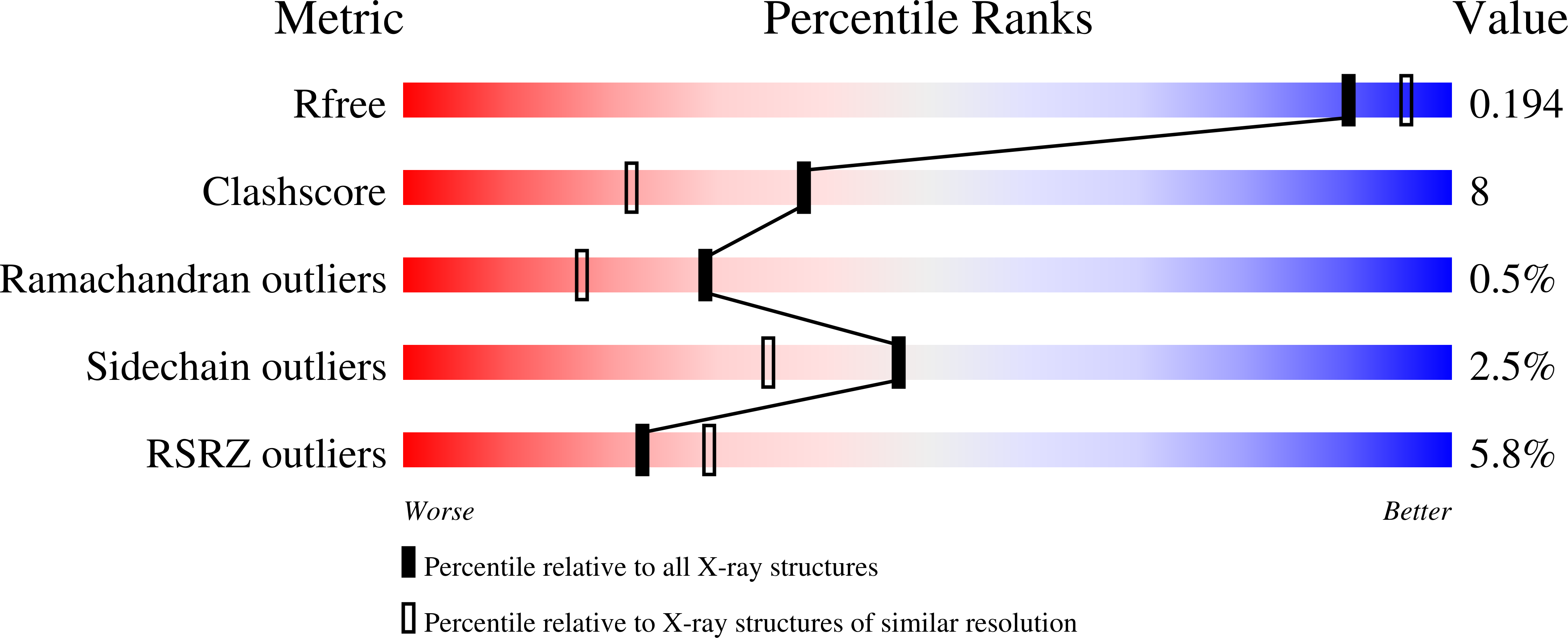
2F8P image
Deposition Date 2005-12-03
Release Date 2006-02-14
Last Version Date 2023-08-30
Entry Detail
PDB ID:
2F8P
Title:
Crystal structure of obelin following Ca2+ triggered bioluminescence suggests neutral coelenteramide as the primary excited state
Biological Source:
Source Organism:
Host Organism:
Method Details:
Experimental Method:
Resolution:
1.93 Å
R-Value Free:
0.22
R-Value Work:
0.19
R-Value Observed:
0.19
Space Group:
P 43
Macromolecular Entities
Polymer Type:polypeptide(L)
Molecule:Obelin
Mutations:S2A
Chain IDs:A
Chain Length:195
Number of Molecules:1
Biological Source:Obelia longissima
Primary Citation

Abstact

The crystal structure at 1.93-A resolution is determined for the Ca2+-discharged obelin containing three bound calcium ions as well as the product of the bioluminescence reaction, coelenteramide. This finding extends the series of available spatial structures of the ligand-dependent conformations of the protein to four, the obelin itself, and those after the bioluminescence reaction with or without bound Ca2+ and/or coelenteramide. Among these structures, global conformational changes are small, typical of the class of "calcium signal modulators" within the EF-hand protein superfamily. Nevertheless, in the active site there are significant repositions of two residues. The His-175 imidazole ring flips becoming almost perpendicular to the original orientation corroborating the crucial importance of this residue for triggering bioluminescence. Tyr-138 hydrogen bonded to the coelenterazine N1-atom in unreacted obelin is moved away from the binding cavity after reaction. However, this Tyr is displaced by a water molecule from within the cavity, which now forms a hydrogen bond to the same atom, the amide N of coelenteramide. From this observation, a reaction scheme is proposed that would result in the neutral coelenteramide as the primary excited state product in photoprotein bioluminescence. From such a higher energy state it is now energetically feasible to account for the shorter wavelength bioluminescence spectra obtained from some photoprotein mutants or to populate the lower energy state of the phenolate anion to yield the blue bioluminescence ordinarily observed from native photoproteins.

Legend

Protein

Chemical

Disease

Primary Citation of related structures