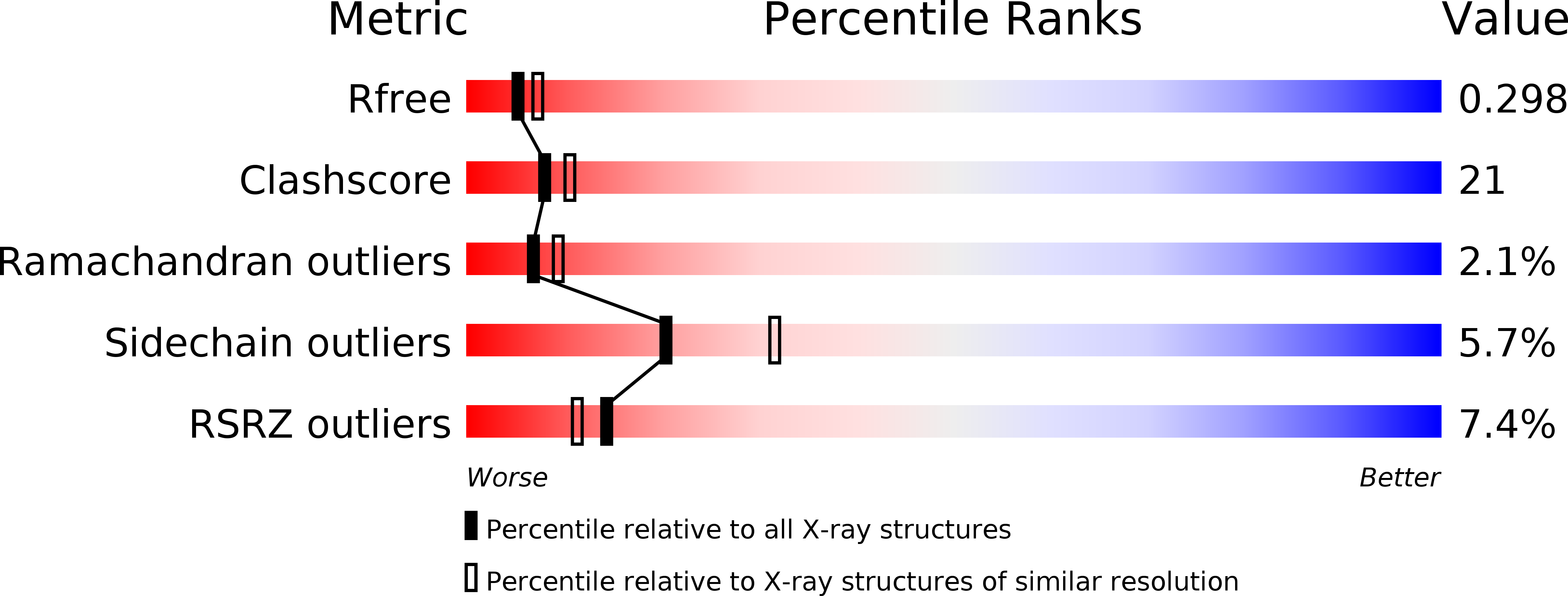
2F86 image
Deposition Date 2005-12-01
Release Date 2006-02-07
Last Version Date 2023-08-30
Entry Detail
PDB ID:
2F86
Keywords:
Title:
The Association Domain of C. elegans CaMKII
Biological Source:
Source Organism:
Host Organism:
Method Details:
Experimental Method:
Resolution:
2.64 Å
R-Value Free:
0.30
R-Value Work:
0.24
R-Value Observed:
0.24
Space Group:
C 2 2 21
Macromolecular Entities
Polymer Type:polypeptide(L)
Molecule:Hypothetical protein K11E8.1d
Gene (Uniprot):unc-43
Chain IDs:A (auth: B), B (auth: D), C (auth: F), D (auth: H), E (auth: J), F (auth: L), G (auth: N)
Chain Length:143
Number of Molecules:7
Biological Source:Caenorhabditis elegans
Primary Citation
Oligomerization states of the association domain and the holoenyzme of Ca2+/CaM kinase II.
Febs J. 273 682 694 (2006)
PMID: 16441656 DOI: 10.1111/j.1742-4658.2005.05088.x

Abstact

Ca2+/calmodulin activated protein kinase II (CaMKII) is an oligomeric protein kinase with a unique holoenyzme architecture. The subunits of CaMKII are bound together into the holoenzyme by the association domain, a C-terminal region of approximately 140 residues in the CaMKII polypeptide. Single particle analyses of electron micrographs have suggested previously that the holoenyzme forms a dodecamer that contains two stacked 6-fold symmetric rings. In contrast, a recent crystal structure of the isolated association domain of mouse CaMKIIalpha has revealed a tetradecameric assembly with two stacked 7-fold symmetric rings. In this study, we have determined the crystal structure of the Caenorhabditis elegans CaMKII association domain and it too forms a tetradecamer. We also show by electron microscopy that in its fully assembled form the CaMKII holoenzyme is a dodecamer but without the kinase domains, either from expression of the isolated association domain in bacteria or following their removal by proteolysis, the association domains form a tetradecamer. We speculate that the holoenzyme is held in its 6-fold symmetric state by the interactions of the N-terminal approximately 1-335 residues and that the removal of this region allows the association domain to convert into a more stable 7-fold symmetric form.

Legend

Protein

Chemical

Disease

Primary Citation of related structures