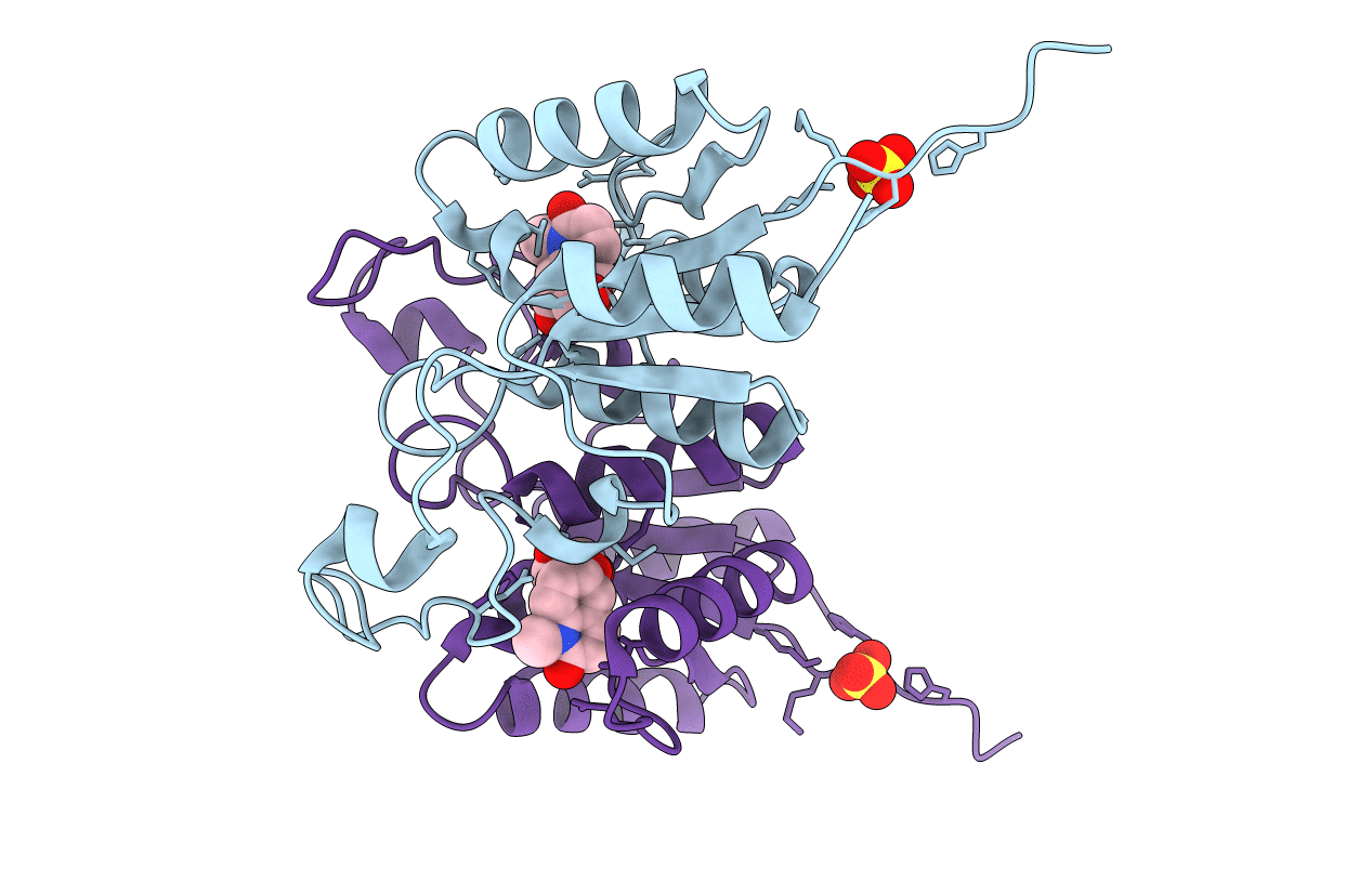
2F64 image
Entry Detail
PDB ID:
2F64
Keywords:
Title:
Crystal structure of Nucleoside 2-deoxyribosyltransferase from Trypanosoma brucei at 1.6 A resolution with 1-METHYLQUINOLIN-2(1H)-ONE bound
Biological Source:
Source Organism:
Host Organism:
PDB Version:
Deposition Date:
2005-11-28
Release Date:
2005-12-06
Method Details:
Experimental Method:
Resolution:
1.60 Å
R-Value Free:
0.18
R-Value Work:
0.16
R-Value Observed:
0.16
Space Group:
C 1 2 1
Macromolecular Entities
Polymer Type:polypeptide(L)
Description:Nucleoside 2-deoxyribosyltransferase
Chain IDs:A, B
Chain Length:161
Number of Molecules:2
Biological Source:Trypanosoma brucei
Modified Residue
Compound ID Chain ID Parent Comp ID Details 2D Image
MSE A MET SELENOMETHIONINE
Primary Citation
Using fragment cocktail crystallography to assist inhibitor design of Trypanosoma brucei nucleoside 2-deoxyribosyltransferase.
J.Med.Chem. 49 5939 5946 (2006)
PMID: 17004709 DOI: 10.1021/jm060429m

Abstact

The 1.8 A resolution de novo structure of nucleoside 2-deoxyribosyltransferase (EC 2.4.2.6) from Trypanosoma brucei (TbNDRT) has been determined by SADa phasing in an unliganded state and several ligand-bound states. This enzyme is important in the salvage pathway of nucleoside recycling. To identify novel lead compounds, we exploited "fragment cocktail soaks". Out of 304 compounds tried in 31 cocktails, four compounds could be identified crystallographically in the active site. In addition, we demonstrated that very short soaks of approximately 10 s are sufficient even for rather hydrophobic ligands to bind in the active site groove, which is promising for the application of similar soaking experiments to less robust crystals of other proteins.

Legend

Protein

Chemical

Disease

Primary Citation of related structures