Deposition Date
2005-11-23
Release Date
2006-03-07
Last Version Date
2024-10-16
Entry Detail
PDB ID:
2F4M
Keywords:
Title:
The Mouse PNGase-HR23 Complex Reveals a Complete Remodulation of the Protein-Protein Interface Compared to its Yeast Orthologs
Biological Source:
Source Organism(s):
Mus musculus (Taxon ID: 10090)
Expression System(s):
Method Details:
Experimental Method:
Resolution:
1.85 Å
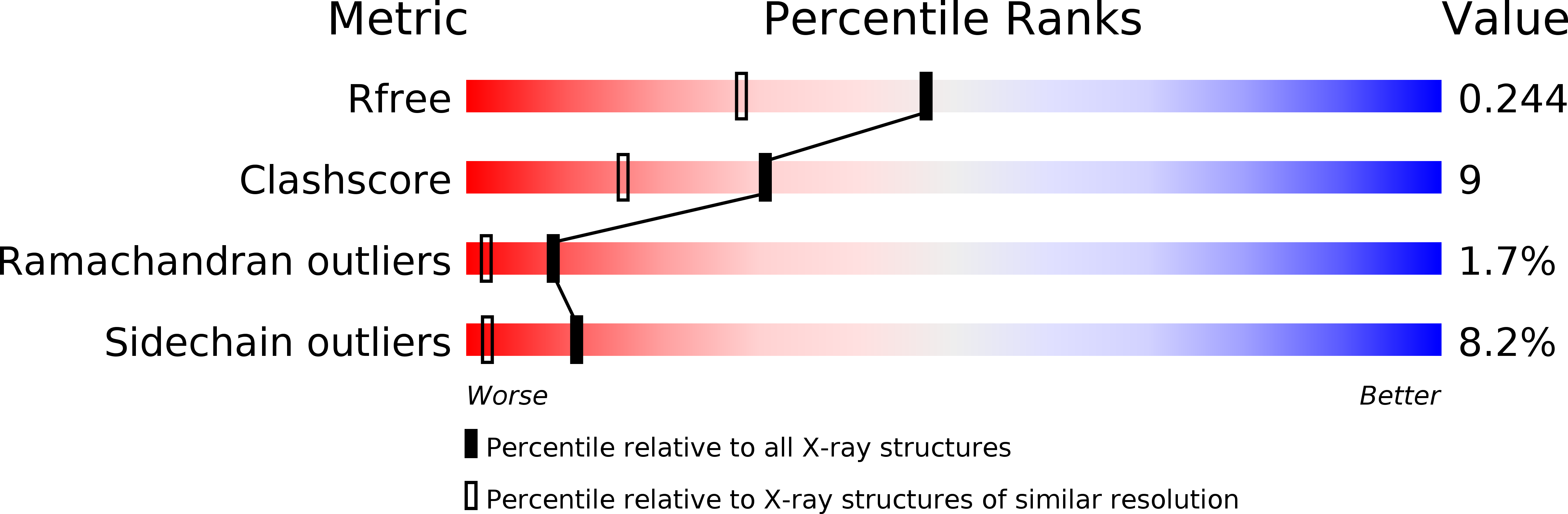
R-Value Free:
0.23
R-Value Work:
0.18
R-Value Observed:
0.18
Space Group:
C 1 2 1