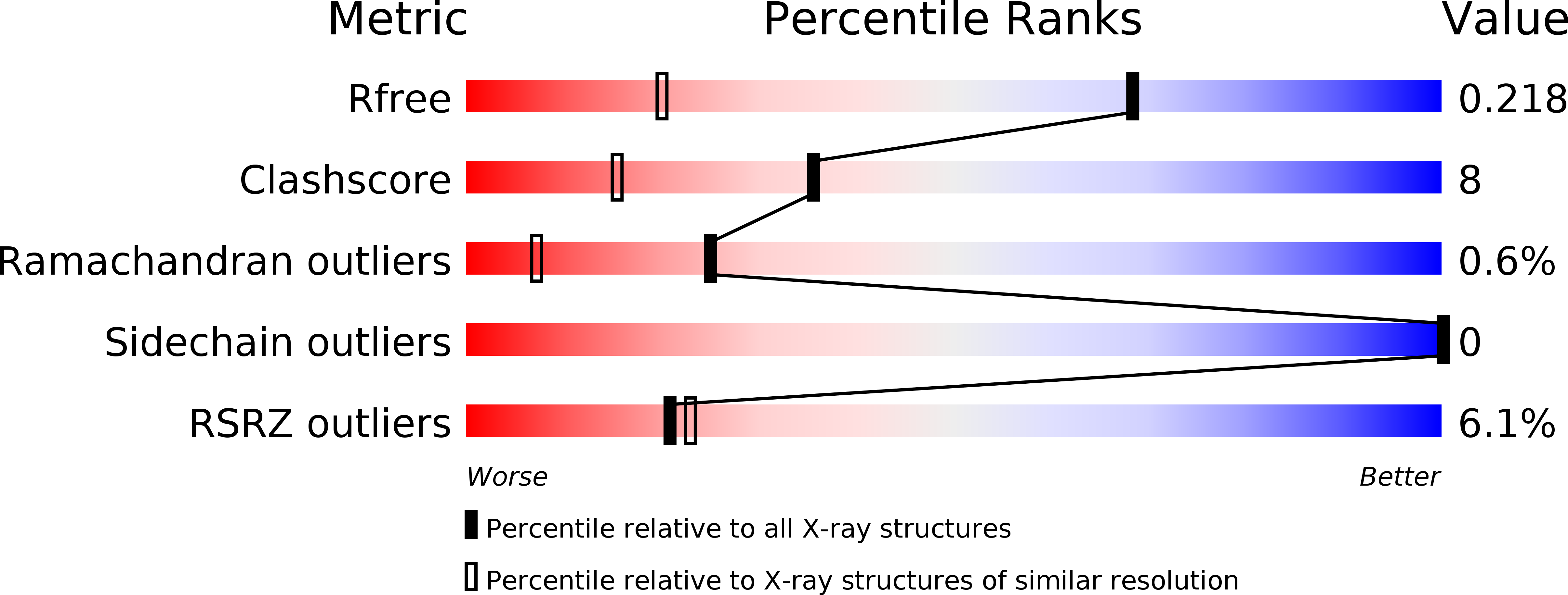
2F3Y image
Deposition Date 2005-11-22
Release Date 2005-12-27
Last Version Date 2024-02-14
Entry Detail
PDB ID:
2F3Y
Title:
Calmodulin/IQ domain complex
Biological Source:
Source Organism:
Homo sapiens (Taxon ID: 9606)
Method Details:
Experimental Method:
Resolution:
1.45 Å
R-Value Free:
0.21
R-Value Work:
0.18
R-Value Observed:
0.18
Space Group:
C 1 2 1
Macromolecular Entities
Structures with similar UniProt ID
Protein Blast
Polymer Type:polypeptide(L)
Molecule:Calmodulin
Chain IDs:A
Chain Length:148
Number of Molecules:1
Biological Source:Homo sapiens
Structures with similar UniProt ID
Protein Blast
Polymer Type:polypeptide(L)
Molecule:Voltage-dependent L-type calcium channel alpha-1C subunit
Chain IDs:B
Chain Length:21
Number of Molecules:1
Biological Source:
Primary Citation
Structure of Calmodulin Bound to the Hydrophobic IQ Domain of the Cardiac Ca(v)1.2 Calcium Channel.
Structure 13 1881 1886 (2005)
PMID: 16338416 DOI: 10.1016/j.str.2005.09.021

Abstact

Ca2+-dependent inactivation (CDI) and facilitation (CDF) of the Ca(v)1.2 Ca2+ channel require calmodulin binding to a putative IQ motif in the carboxy-terminal tail of the pore-forming subunit. We present the 1.45 A crystal structure of Ca2+-calmodulin bound to a 21 residue peptide corresponding to the IQ domain of Ca(v)1.2. This structure shows that parallel binding of calmodulin to the IQ domain is governed by hydrophobic interactions. Mutations of residues I1672 and Q1673 in the peptide to alanines, which abolish CDI but not CDF in the channel, do not greatly alter the structure. Both lobes of Ca2+-saturated CaM bind to the IQ peptide but isoleucine 1672, thought to form an intramolecular interaction that drives CDI, is buried. These findings suggest that this structure could represent the conformation that calmodulin assumes in CDF.

Legend

Protein

Chemical

Disease

Primary Citation of related structures
Feedback Form
Name
Email
Institute
Feedback