Deposition Date
2005-11-22
Release Date
2006-05-30
Last Version Date
2023-08-23
Entry Detail
PDB ID:
2F3T
Keywords:
Title:
Crystal Structure Of E.coli Guanylate Kinase In Complex With Ganciclovir monophosphate
Biological Source:
Source Organism(s):
Escherichia coli (Taxon ID: 562)
Expression System(s):
Method Details:
Experimental Method:
Resolution:
3.16 Å
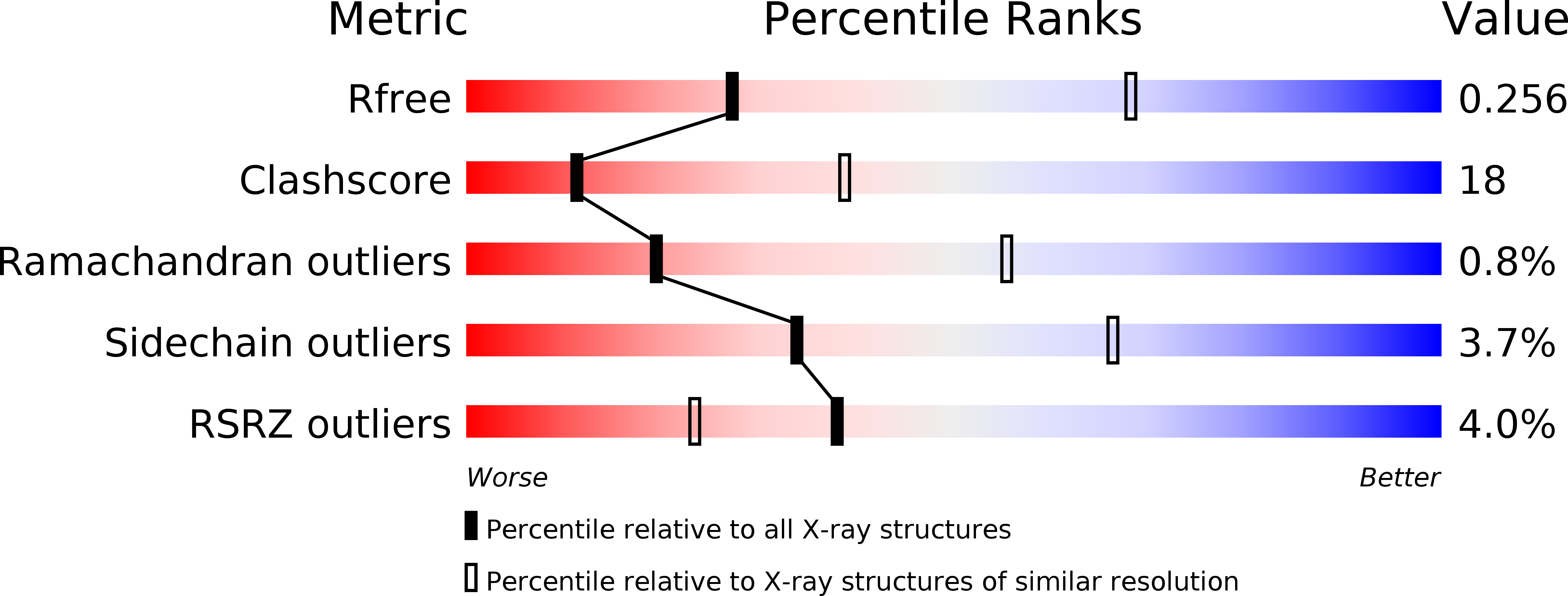
R-Value Free:
0.29
R-Value Work:
0.26
R-Value Observed:
0.26
Space Group:
P 41 21 2