2F3O image
Deposition Date 2005-11-22
Release Date 2006-03-07
Last Version Date 2023-08-23
Entry Detail
PDB ID:
2F3O
Title:
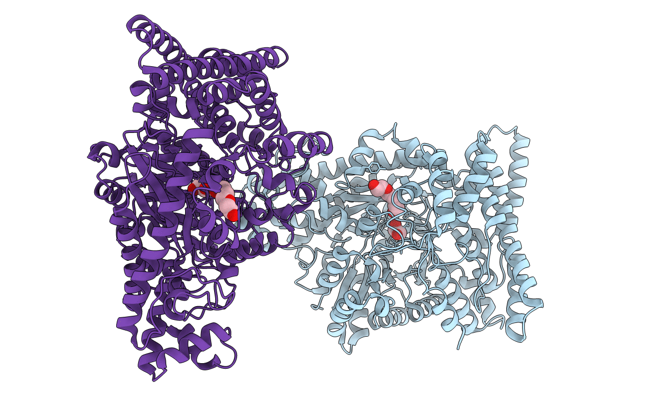
Crystal Structure of a glycyl radical enzyme from Archaeoglobus fulgidus
Biological Source:
Source Organism:
Host Organism:
Method Details:
Experimental Method:
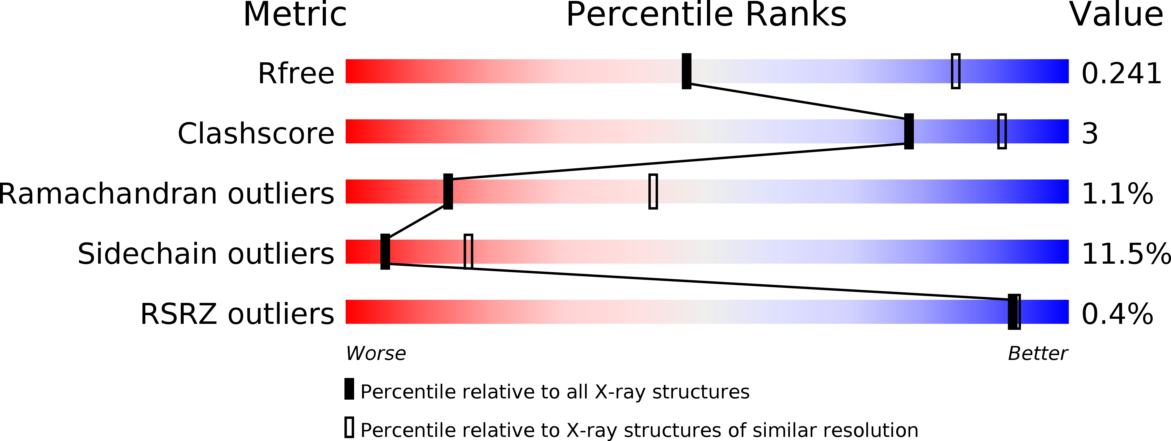
Resolution:
2.90 Å
R-Value Free:
0.24
R-Value Work:
0.19
R-Value Observed:
0.20
Space Group:
C 2 2 2
Macromolecular Entities
Polymer Type:polypeptide(L)
Molecule:pyruvate formate-lyase 2
Gene (Uniprot):AF_1449
Chain IDs:A, B
Chain Length:776
Number of Molecules:2
Biological Source:Archaeoglobus fulgidus
Primary Citation
Crystal structure of a glycyl radical enzyme from Archaeoglobus fulgidus
J.Mol.Biol. 357 221 235 (2006)
PMID: 16414072 DOI: 10.1016/j.jmb.2005.12.049

Abstact

We have solved the crystal structure of a PFL2 from Archaeglobus fulgidus at 2.9 A resolution. Of the three previously solved enzyme structures of glycyl radical enzymes, pyruvate formate lyase (PFL), anaerobic ribonucleotide reductase and glycerol dehydratase (GD), the last one is clearly most similar to PFL2. We observed electron density in the active site of PFL2, which we modelled as glycerol. The orientation of the glycerol is different from that in GD, and changes in the active site indicate that the actual substrate of PFL2 is bigger than a glycerol molecule, but sequence and structural homology suggest that PFL2 may be a dehydratase. Crystal packing, solution X-ray scattering and ultracentrifugation experiments show that PFL2 is tetrameric, unlike other glycyl radical enzymes. A.fulgidus is a hyperthermophile and PFL2 appears to be stabilized by several factors including an increased number of ion pairs, differences in buried charges, a truncated N terminus, anchoring of loops and N terminus via salt-bridges, changes in the oligomeric interface and perhaps also the higher oligomerization state of the protein.

Legend

Protein

Chemical

Disease

Primary Citation of related structures