Deposition Date
2005-11-18
Release Date
2006-10-31
Last Version Date
2023-08-23
Entry Detail
PDB ID:
2F38
Keywords:
Title:
Crystal structure of prostaglandin F synathase containing bimatoprost
Biological Source:
Source Organism(s):
Homo sapiens (Taxon ID: 9606)
Expression System(s):
Method Details:
Experimental Method:
Resolution:
2.00 Å
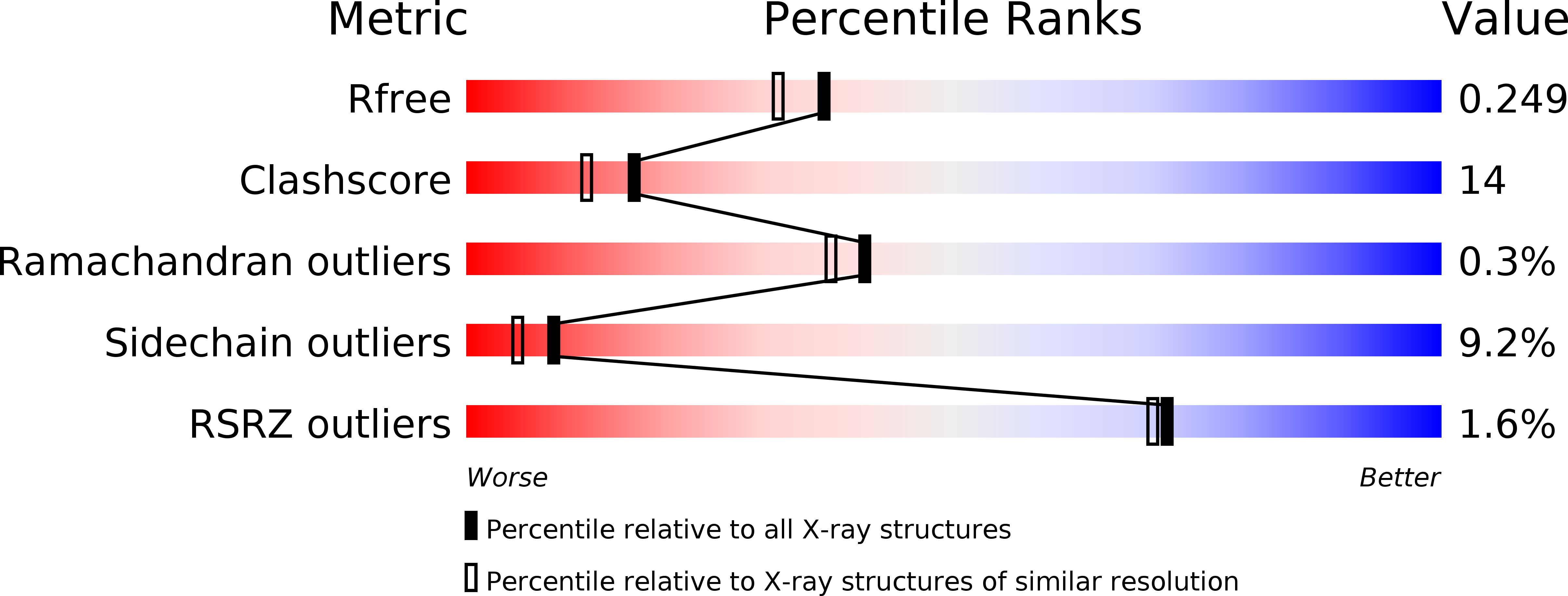
R-Value Free:
0.28
R-Value Work:
0.22
R-Value Observed:
0.22
Space Group:
P 1 21 1