2F37 image
Deposition Date 2005-11-18
Release Date 2006-10-31
Last Version Date 2024-11-13
Entry Detail
PDB ID:
2F37
Title:
Crystal structure of the ankyrin repeat domain of human TRPV2
Biological Source:
Source Organism:
Homo sapiens (Taxon ID: 9606)
Host Organism:
Method Details:
Experimental Method:
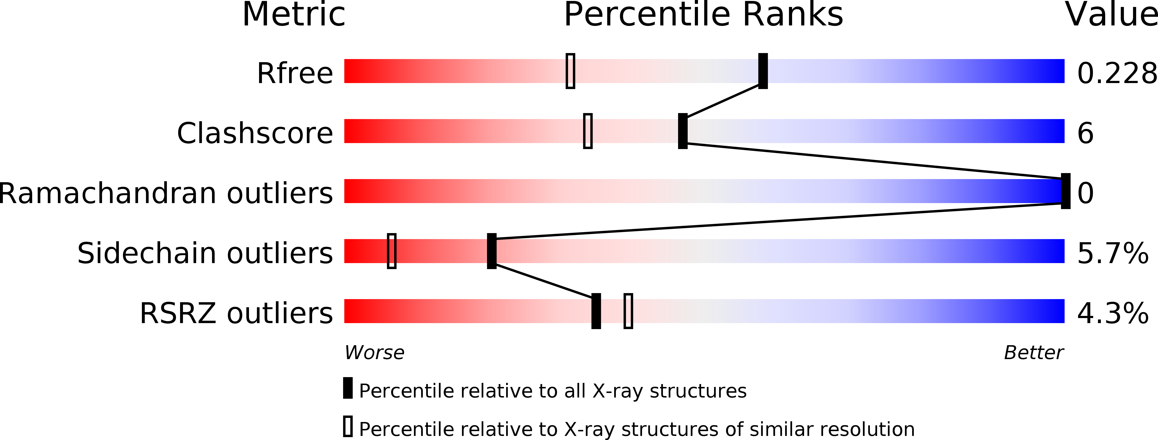
Resolution:
1.70 Å
R-Value Free:
0.22
R-Value Work:
0.20
R-Value Observed:
0.20
Space Group:
P 61 2 2
Macromolecular Entities
Polymer Type:polypeptide(L)
Molecule:Transient receptor potential cation channel subfamily V member 2
Gene (Uniprot):TRPV2
Chain IDs:A, B
Chain Length:251
Number of Molecules:2
Biological Source:Homo sapiens
Modified Residue
Compound ID Chain ID Parent Comp ID Details 2D Image
MSE A MET SELENOMETHIONINE
Ligand Molecules
Primary Citation
Crystal structure of the human TRPV2 channel ankyrin repeat domain.
Protein Sci. 15 2201 2206 (2006)
PMID: 16882997 DOI: 10.1110/ps.062357206

Abstact

TRPV channels are important polymodal integrators of noxious stimuli mediating thermosensation and nociception. An ankyrin repeat domain (ARD), which is a common protein-protein recognition domain, is conserved in the N-terminal intracellular domain of all TRPV channels and predicted to contain three to four ankyrin repeats. Here we report the first structure from the TRPV channel subfamily, a 1.7 A resolution crystal structure of the human TRPV2 ARD. Our crystal structure reveals a six ankyrin repeat stack with multiple insertions in each repeat generating several unique features compared with a canonical ARD. The surface typically used for ligand recognition, the ankyrin groove, contains extended loops with an exposed hydrophobic patch and a prominent kink resulting from a large rotational shift of the last two repeats. The TRPV2 ARD provides the first structural insight into a domain that coordinates nociceptive sensory transduction and is likely to be a prototype for other TRPV channel ARDs.

Legend

Protein

Chemical

Disease

Primary Citation of related structures