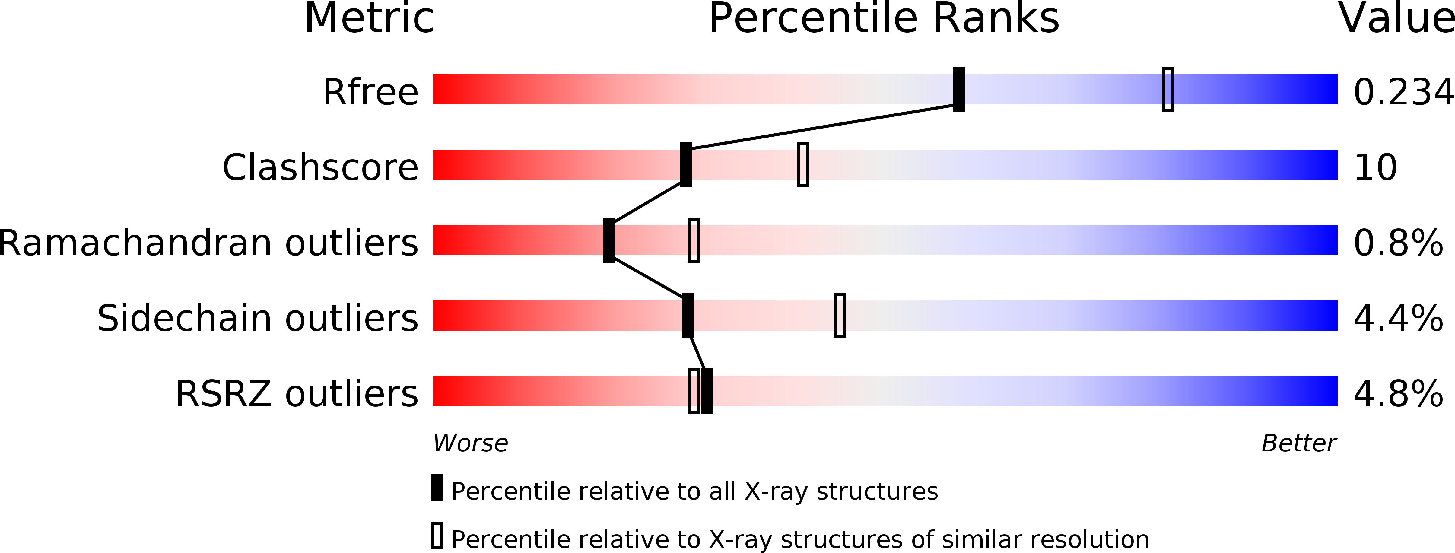
2F2U image
Entry Detail
PDB ID:
2F2U
Keywords:
Title:
crystal structure of the Rho-kinase kinase domain
Biological Source:
Source Organism:
Host Organism:
PDB Version:
Deposition Date:
2005-11-18
Release Date:
2006-04-25
Method Details:
Experimental Method:
Resolution:
2.40 Å
R-Value Free:
0.23
R-Value Work:
0.19
R-Value Observed:
0.19
Space Group:
P 41 21 2
Macromolecular Entities
Polymer Type:polypeptide(L)
Description:Rho-associated protein kinase 2
Chain IDs:A, B
Chain Length:402
Number of Molecules:2
Biological Source:Bos taurus
Ligand Molecules
Primary Citation
Molecular mechanism for the regulation of rho-kinase by dimerization and its inhibition by fasudil
Structure 14 589 600 (2006)
PMID: 16531242 DOI: 10.1016/j.str.2005.11.024

Abstact

Rho-kinase is a key regulator of cytoskeletal events and a promising drug target in the treatment of vascular diseases and neurological disorders. Unlike other protein kinases, Rho-kinase requires both N- and C-terminal extension segments outside the kinase domain for activity, although the details of this requirement have been elusive. The crystal structure of an active Rho-kinase fragment containing the kinase domain and both the extensions revealed a head-to-head homodimer through the N-terminal extension forming a helix bundle that structurally integrates the C-terminal extension. This structural organization enables binding of the C-terminal hydrophobic motif to the N-terminal lobe, which defines the correct disposition of helix alphaC that is important for the catalytic activity. The bound inhibitor fasudil significantly alters the conformation and, consequently, the mode of interaction with the catalytic cleft that contains local structural changes. Thus, both kinase and drug conformational pliability and stability confer selectivity.

Legend

Protein

Chemical

Disease

Primary Citation of related structures