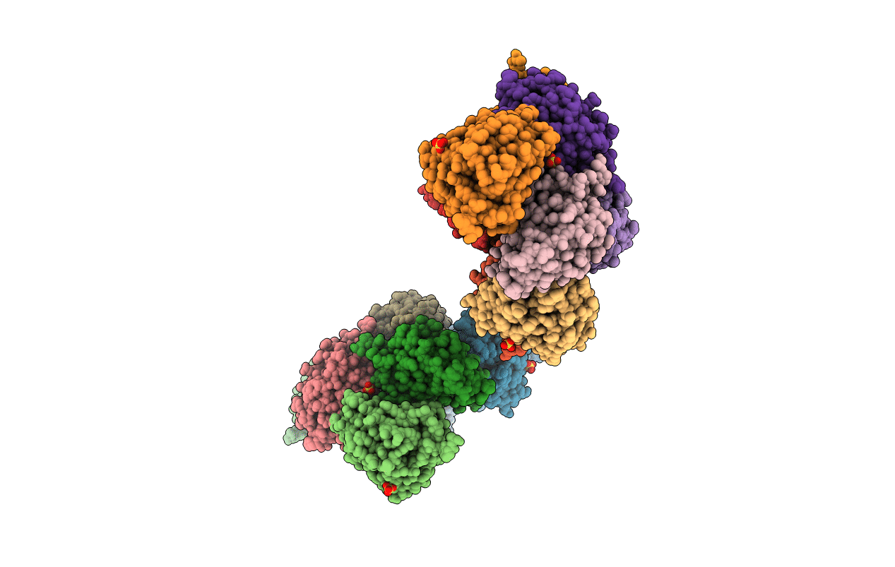
2F1D image
Deposition Date 2005-11-14
Release Date 2006-01-24
Last Version Date 2023-08-23
Entry Detail
PDB ID:
2F1D
Keywords:
Title:
X-Ray Structure of imidazoleglycerol-phosphate dehydratase
Biological Source:
Source Organism:
Method Details:
Experimental Method:
Resolution:
3.00 Å
R-Value Free:
0.28
R-Value Work:
0.24
R-Value Observed:
0.24
Space Group:
H 3
Macromolecular Entities
Polymer Type:polypeptide(L)
Molecule:Imidazoleglycerol-phosphate dehydratase 1
Gene (Uniprot):HISN5A
Chain IDs:A, B, C, D, E, F, G, H, I, J, K, L, M, N, O, P
Chain Length:207
Number of Molecules:16
Biological Source:Arabidopsis thaliana
Primary Citation
Structure and mechanism of imidazoleglycerol-phosphate dehydratase.
Structure 13 1809 1817 (2005)
PMID: 16338409 DOI: 10.1016/j.str.2005.08.012

Abstact

The structure of A. thaliana imidazoleglycerol-phosphate dehydratase, an enzyme of histidine biosynthesis and a target for the triazole phosphonate herbicides, has been determined to 3.0 A resolution. The structure is composed of 24 identical subunits arranged in 432 symmetry and shows how the formation of a novel dimanganese cluster is crucial to the assembly of the active 24-mer from an inactive trimeric precursor and to the formation of the active site of the enzyme. Molecular modeling suggests that the substrate is bound to the manganese cluster as an imidazolate moiety that subsequently collapses to yield a diazafulvene intermediate. The mode of imidazolate recognition exploits pseudosymmetry at the active site arising from a combination of the assembly of the particle and the pseudosymmetry present in each subunit as a result of gene duplication. This provides an intriguing example of the role of evolution in the design of Nature's catalysts.

Legend

Protein

Chemical

Disease

Primary Citation of related structures