Deposition Date
1992-05-27
Release Date
1992-10-15
Last Version Date
2024-11-13
Entry Detail
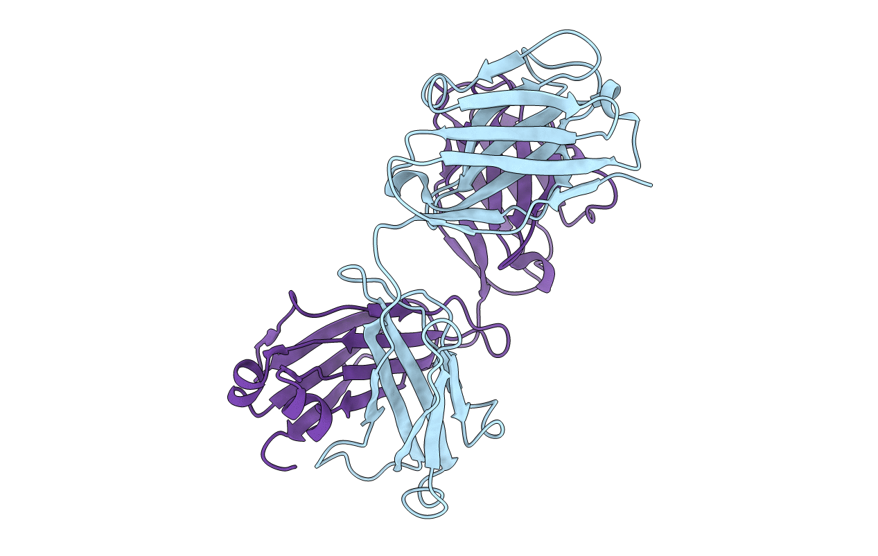
PDB ID:
2F19
Keywords:
Title:
THREE-DIMENSIONAL STRUCTURE OF TWO CRYSTAL FORMS OF FAB R19.9, FROM A MONOCLONAL ANTI-ARSONATE ANTIBODY
Biological Source:
Source Organism(s):
Mus musculus (Taxon ID: 10090)
Method Details: