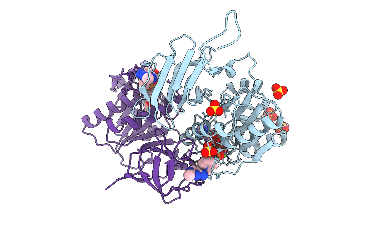
2F17 image
Deposition Date 2005-11-14
Release Date 2005-11-29
Last Version Date 2023-08-23
Entry Detail
PDB ID:
2F17
Keywords:
Title:
Mouse Thiamin Pyrophosphokinase in a Ternary Complex with Pyrithiamin Pyrophosphate and AMP at 2.5 angstrom
Biological Source:
Source Organism:
Mus musculus (Taxon ID: 10090)
Host Organism:
Method Details:
Experimental Method:
Resolution:
2.50 Å
R-Value Free:
0.25
R-Value Work:
0.23
R-Value Observed:
0.24
Space Group:
P 31 2 1
Macromolecular Entities
Structures with similar UniProt ID
Protein Blast
Polymer Type:polypeptide(L)
Molecule:Thiamin pyrophosphokinase 1
Gene (Uniprot):Tpk1
Chain IDs:A, B
Chain Length:263
Number of Molecules:2
Biological Source:Mus musculus
Primary Citation
Pyrithiamine as a substrate for thiamine pyrophosphokinase
J.Biol.Chem. 281 6601 6607 (2006)
PMID: 16365036 DOI: 10.1074/jbc.M510951200

Abstact

Thiamine pyrophosphokinase transfers a pyrophosphate group from a nucleoside triphosphate, such as ATP, to the hydroxyl group of thiamine to produce thiamine pyrophosphate. Deficiencies in thiamine can result in the development of the neurological disorder Wernicke-Korsakoff Syndrome as well as the potentially fatal cardiovascular disease wet beriberi. Pyrithiamine is an inhibitor of thiamine metabolism that induces neurological symptoms similar to that of Wernicke-Korsakoff Syndrome in animals. However, the mechanism by which pyrithiamine interferes with cellular thiamine phosphoester homeostasis is not entirely clear. We used kinetic assays coupled with mass spectrometry of the reaction products and x-ray crystallography of an equilibrium reaction mixture of thiamine pyrophosphokinase, pyrithiamine, and Mg2+/ATP to elucidate the mechanism by which pyrithiamine inhibits the enzymatic production of thiamine pyrophosphate. Three lines of evidence support the ability of thiamine pyrophosphokinase to form pyrithiamine pyrophosphate. First, a coupled enzyme assay clearly demonstrated the ability of thiamine pyrophosphokinase to produce AMP when pyrithiamine was used as substrate. Second, an analysis of the reaction mixture by mass spectrometry directly identified pyrithiamine pyrophosphate in the reaction mixture. Last, the structure of thiamine pyrophosphokinase crystallized from an equilibrium substrate/product mixture shows clear electron density for pyrithiamine pyrophosphate bound in the enzyme active site. This structure also provides the first clear picture of the binding pocket for the nucleoside triphosphate and permits the first detailed understanding of the catalytic requirements for catalysis in this enzyme.

Legend

Protein

Chemical

Disease

Primary Citation of related structures