2F14 image
Deposition Date 2005-11-14
Release Date 2006-10-24
Last Version Date 2023-08-23
Entry Detail
PDB ID:
2F14
Keywords:
Title:
Tne Crystal Structure of the Human Carbonic Anhydrase II in Complex with a Fluorescent Inhibitor
Biological Source:
Source Organism:
Homo sapiens (Taxon ID: 9606)
Method Details:
Experimental Method:
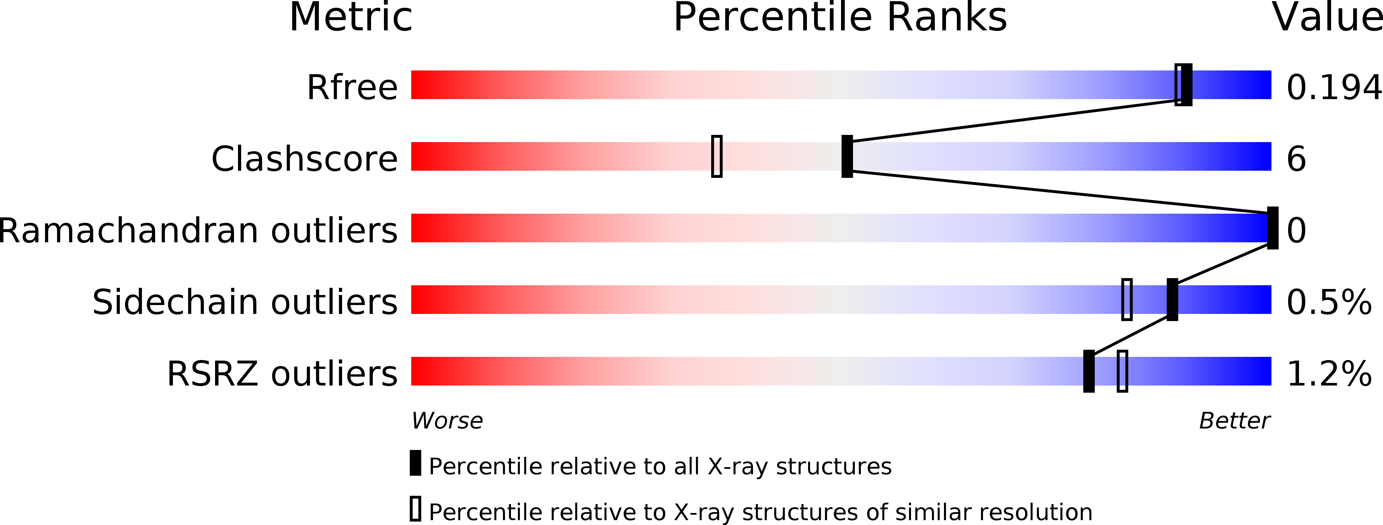
Resolution:
1.71 Å
R-Value Free:
0.20
R-Value Work:
0.17
Space Group:
P 1 21 1
Macromolecular Entities
Polymer Type:polypeptide(L)
Molecule:Carbonic anhydrase 2
Gene (Uniprot):CA2
Chain IDs:A
Chain Length:259
Number of Molecules:1
Biological Source:Homo sapiens
Primary Citation
Carbonic anhydrase inhibitors: X-ray and molecular modeling study for the interaction of a fluorescent antitumor sulfonamide with isozyme II and IX.
J.Am.Chem.Soc. 128 8329 8335 (2006)
PMID: 16787097 DOI: 10.1021/ja061574s

Abstact

The X-ray crystal structure of the fluorescent antitumor sulfonamide carbonic anhydrase (CA, EC, 4.2.1.1) inhibitor (4-sulfamoylphenylethyl)thioureido fluorescein (1) in complex with the cytosolic isoform hCA II is reported, together with a modeling study of the adduct of 1 with the tumor-associated isoform hCA IX. Its binding to hCA II is similar to that of other benzesulfonamides, with the ionized sulfonamide coordinated to the Zn2+ ion within the enzyme active site, and also participating in a network of hydrogen bonds with residues Thr199 and Glu106. The scaffold of 1 did not establish polar interactions within the enzyme active site but made hydrophobic contacts (<4.5 A) with Gln92, Val121, Phe131, Val135, Leu198, Thr199, Thr200, and Pro202. The substituted 3-carboxy-amino-phenyl functionality was at van der Waals distance from Phe131, Gly132, and Val135. The bulky tricyclic fluorescein moiety was located at the rim of the active site, on the protein surface, and strongly interacted with the alpha-helix formed by residues Asp130-Val135. All these interactions were preserved in the hCA IX-1 adduct, but the carbonyl moiety of the fluorescein tail of 1 participates in a strong hydrogen bond with the guanidine moiety of Arg130, an amino acid characteristic of the hCA IX active site. This may account for the roughly 2 times higher affinity of 1 for hCA IX over hCA II and may explain why in vivo the compound specifically accumulates only in hypoxic tumors overexpressing CA IX and not in the normal tissues. The compound is in clinical studies as an imaging tool for acute hypoxic tumors.

Legend

Protein

Chemical

Disease

Primary Citation of related structures