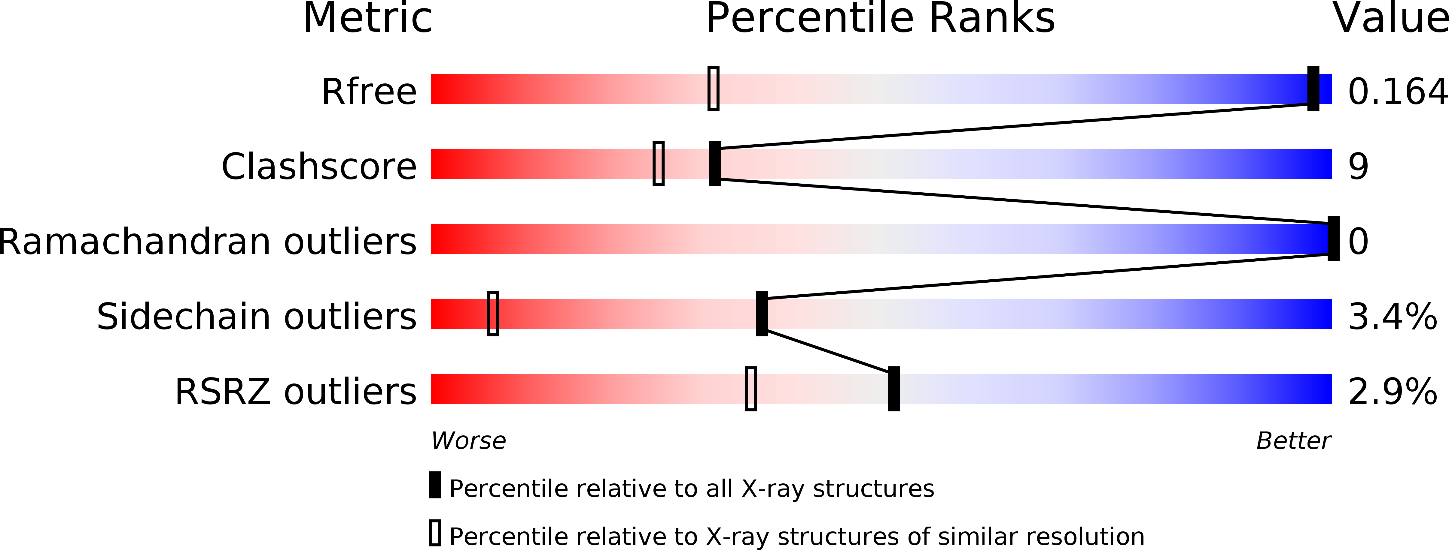
2F01 image
Deposition Date 2005-11-10
Release Date 2005-11-29
Last Version Date 2023-08-23
Entry Detail
PDB ID:
2F01
Title:
Epi-biotin complex with core streptavidin
Biological Source:
Source Organism:
Host Organism:
Method Details:
Experimental Method:
Resolution:
0.85 Å
R-Value Free:
0.17
R-Value Work:
0.15
R-Value Observed:
0.15
Space Group:
I 2 2 2
Macromolecular Entities
Polymer Type:polypeptide(L)
Molecule:Streptavidin
Chain IDs:A, B
Chain Length:127
Number of Molecules:2
Biological Source:Streptomyces avidinii
Primary Citation
The high-resolution structure of (+)-epi-biotin bound to streptavidin.
Acta Crystallogr.,Sect.D 62 576 581 (2006)
PMID: 16699183 DOI: 10.1107/S0907444906011887

Abstact

(+)-Epi-biotin differs from (+)-biotin in the configuration of the chiral center at atom C2. This could lead to a difference in the mode of binding of (+)-epi-biotin to streptavidin, a natural protein receptor for (+)-biotin. Diffraction data were collected to a maximum of 0.85 Angstrom resolution for structural analysis of the complex of streptavidin with a sample of (+)-epi-biotin and refinement was carried out at both 1.0 and 0.85 Angstrom resolution. The structure determination shows a superposition of two ligands in the binding site, (+)-biotin and (+)-epi-biotin. The molecules overlap in the model for the complex except for the position of S1 in the tetrahydrothiophene ring. Differences in the conformation of the ring permits binding of each molecule to streptavidin with little observable difference in the protein structures at this high resolution.

Legend

Protein

Chemical

Disease

Primary Citation of related structures