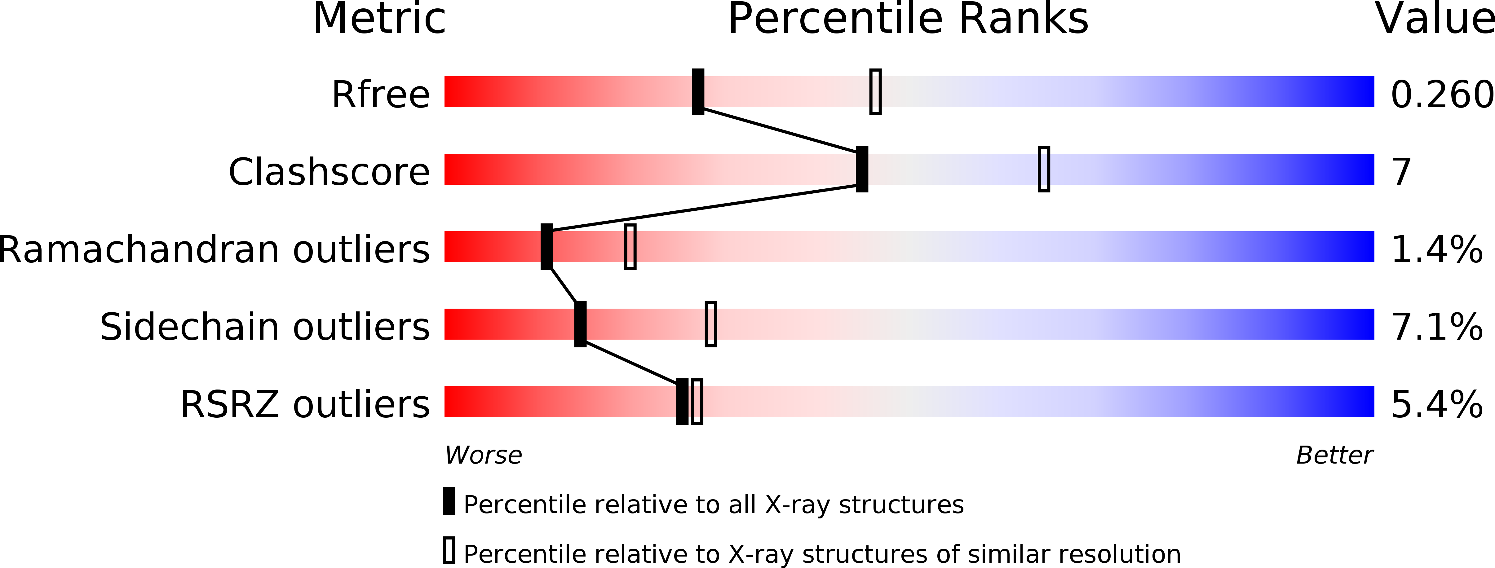
2F00 image
Deposition Date 2005-11-10
Release Date 2006-10-24
Last Version Date 2024-11-20
Entry Detail
PDB ID:
2F00
Keywords:
Title:
Escherichia coli MurC
Biological Source:
Source Organism:
Escherichia coli (Taxon ID: 562)
Host Organism:
Method Details:
Experimental Method:
Resolution:
2.50 Å
R-Value Free:
0.26
R-Value Work:
0.21
Space Group:
P 21 21 21
Macromolecular Entities
Polymer Type:polypeptide(L)
Molecule:UDP-N-acetylmuramate--L-alanine ligase
Gene (Uniprot):murC
Chain IDs:A, B
Chain Length:491
Number of Molecules:2
Biological Source:Escherichia coli
Modified Residue
Compound ID Chain ID Parent Comp ID Details 2D Image
MSE A MET SELENOMETHIONINE
Ligand Molecules
Primary Citation
Structure of Escherichia coliUDP-N-acetylmuramoyl:L-alanine ligase (MurC).
Acta Crystallogr.,Sect.D 62 1466 1474 (2006)
PMID: 17139082 DOI: 10.1107/S0907444906038376

Abstact

The bacterial cell wall provides essential protection from the external environment and confers strength and rigidity to counteract internal osmotic pressure. Without this layer the cell would be easily ruptured and it is for this reason that biosynthetic pathways leading to the formation of peptidoglycan have for many years been a prime target for effective antibiotics. Central to this pathway are four similar ligase enzymes which add peptide groups to glycan moieties. As part of a program to better understand the structure-function relationships in these four enzymes, the crystal structure of Escherichia coli UDP-N-acetylmuramoyl:L-alanine ligase (MurC) has been determined to 2.6 A resolution. The structure was solved by multiwavelength anomalous diffraction methods from a single selenomethionine-substituted crystal and refined to a crystallographic R factor of 0.212 (R(free) = 0.259). The enzyme has a modular multi-domain structure very similar to those of other members of the mur family of ATP-dependent amide-bond ligases. Detailed comparison of these four enzymes shows that considerable conformational changes are possible. These changes, together with the recruitment of two different N-terminal domains, allow this family of enzymes to bind a substrate which is identical at one end and at the other has the growing peptide tail which will ultimately become part of the rigid bacterial cell wall. Comparison of the E. coli and Haemophilus influenzae structures and analysis of the sequences of known MurC enzymes indicate the presence of a ;dimerization' motif in almost 50% of the MurC enzymes and points to a highly conserved loop in domain 3 that may play a key role in amino-acid ligand specificity.

Legend

Protein

Chemical

Disease

Primary Citation of related structures