
Deposition Date
1997-07-25
Release Date
1997-12-03
Last Version Date
2024-05-29
Entry Detail
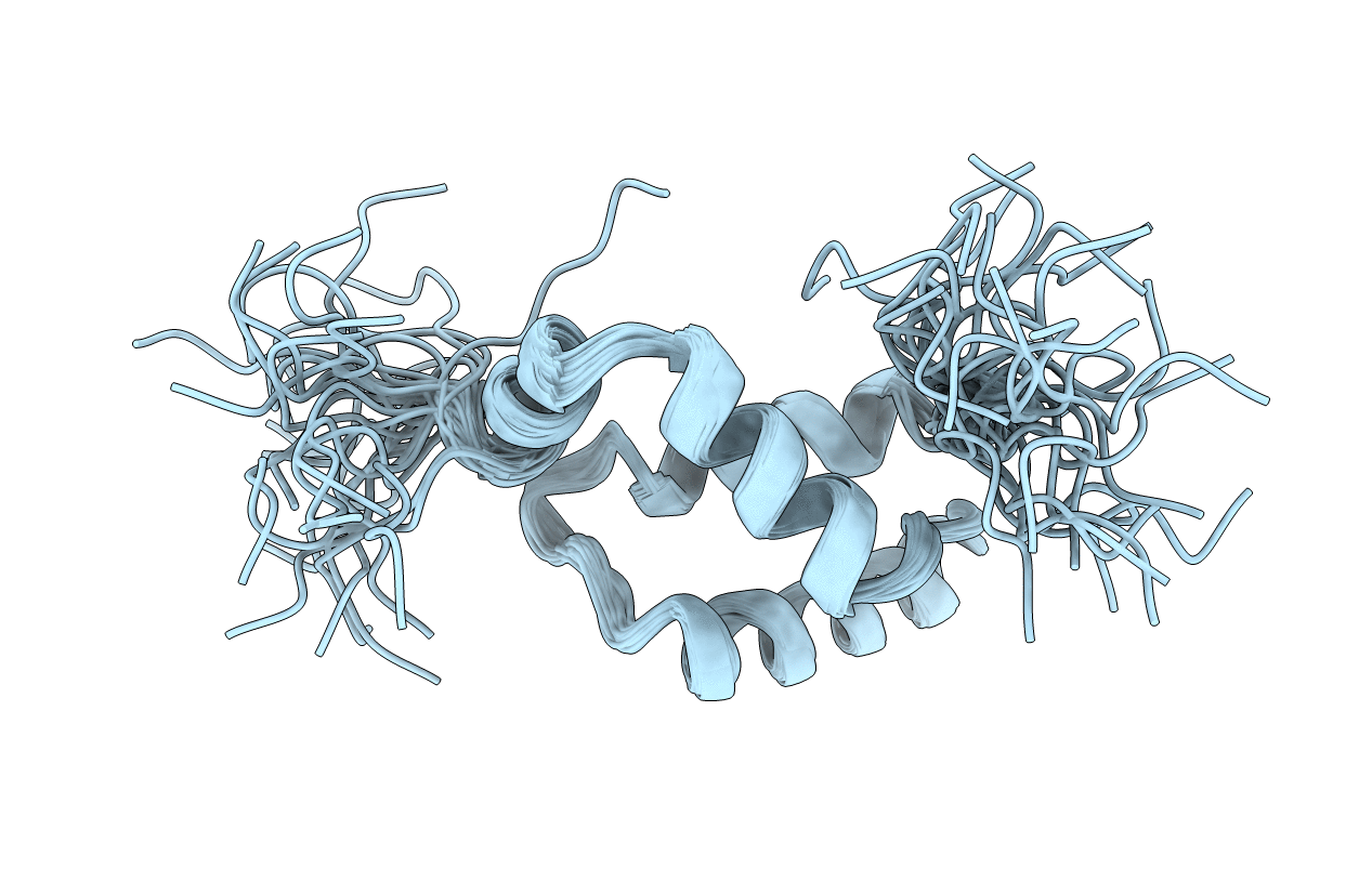
PDB ID:
2EZI
Keywords:
Title:
SOLUTION NMR STRUCTURE OF THE IGAMMA SUBDOMAIN OF THE MU END DNA BINDING DOMAIN OF MU PHAGE TRANSPOSASE, 30 STRUCTURES
Biological Source:
Source Organism:
Enterobacteria phage Mu (Taxon ID: 10677)
Method Details:
Experimental Method:
Conformers Submitted:
30
