2EX6 image
Entry Detail
PDB ID:
2EX6
Keywords:
Title:
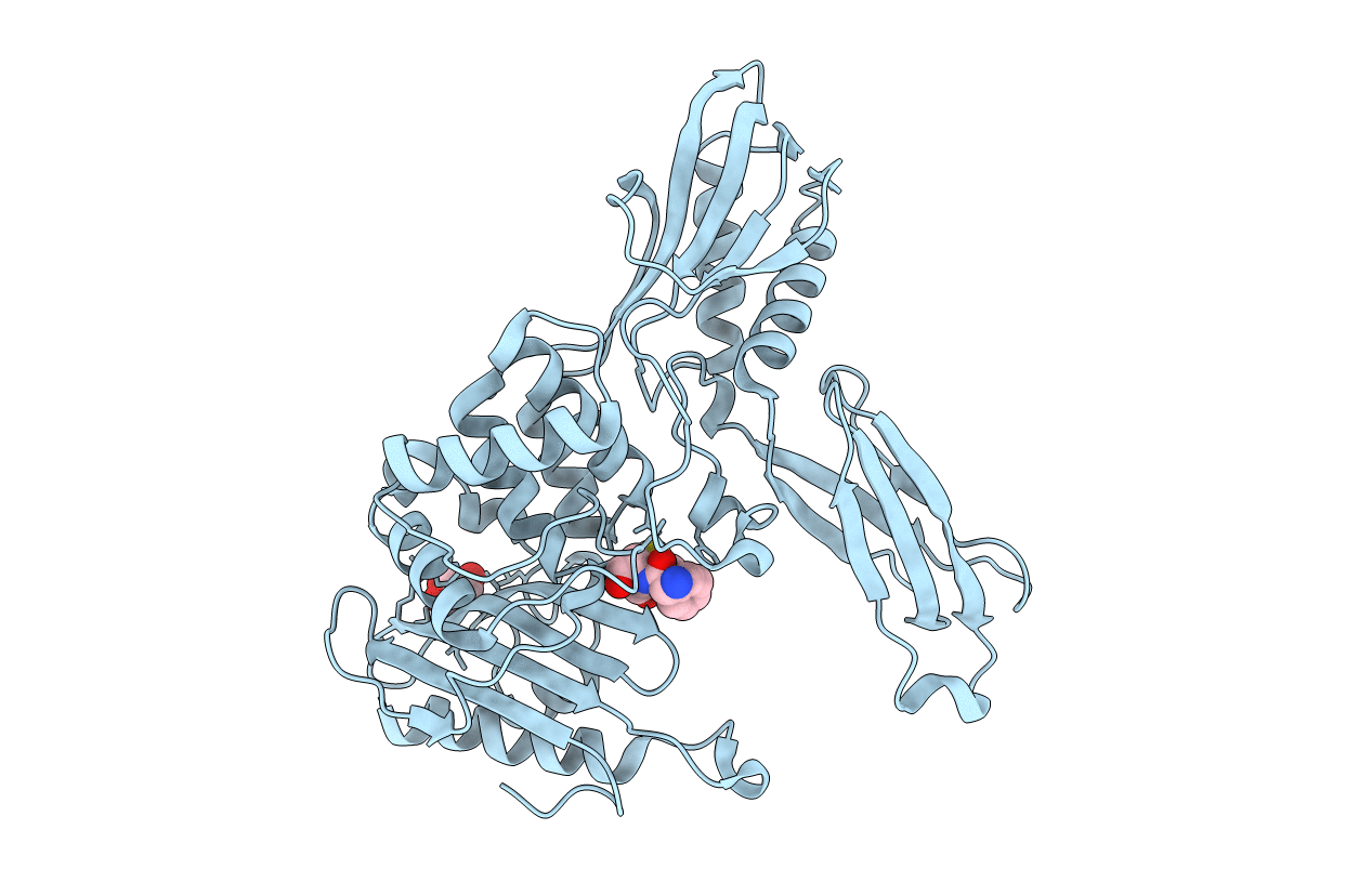
Crystal structure of penicillin binding protein 4 (dacB) from Escherichia coli, complexed with ampicillin
Biological Source:
Source Organism:
PDB Version:
Deposition Date:
2005-11-08
Release Date:
2006-06-13
Method Details:
Experimental Method:
Resolution:
1.60 Å
R-Value Free:
0.24
R-Value Work:
0.21
R-Value Observed:
0.21
Space Group:
P 41 21 2
Macromolecular Entities
Polymer Type:polypeptide(L)
Description:Penicillin-binding protein 4
Mutations:D261Y
Chain IDs:A
Chain Length:458
Number of Molecules:1
Biological Source:Escherichia coli
Primary Citation
Crystal structure of penicillin binding protein 4 (dacB) from Escherichia coli, both in the native form and covalently linked to various antibiotics
Biochemistry 45 783 792 (2006)
PMID: 16411754 DOI: 10.1021/bi051533t

Abstact

The crystal structure of penicillin binding protein 4 (PBP4) from Escherichia coli, which has both DD-endopeptidase and DD-carboxypeptidase activity, is presented. PBP4 is one of 12 penicillin binding proteins in E. coli involved in the synthesis and maintenance of the cell wall. The model contains a penicillin binding domain similar to known structures, but includes a large insertion which folds into domains with unique folds. The structures of the protein covalently attached to five different antibiotics presented here show the active site residues are unmoved compared to the apoprotein, but nearby surface loops and helices are displaced in some cases. The altered geometry of conserved active site residues compared with those of other PBPs suggests a possible cause for the slow deacylation rate of PBP4.

Legend

Protein

Chemical

Disease

Primary Citation of related structures