
Deposition Date
2005-11-07
Release Date
2006-02-28
Last Version Date
2023-08-23
Entry Detail
PDB ID:
2EX1
Keywords:
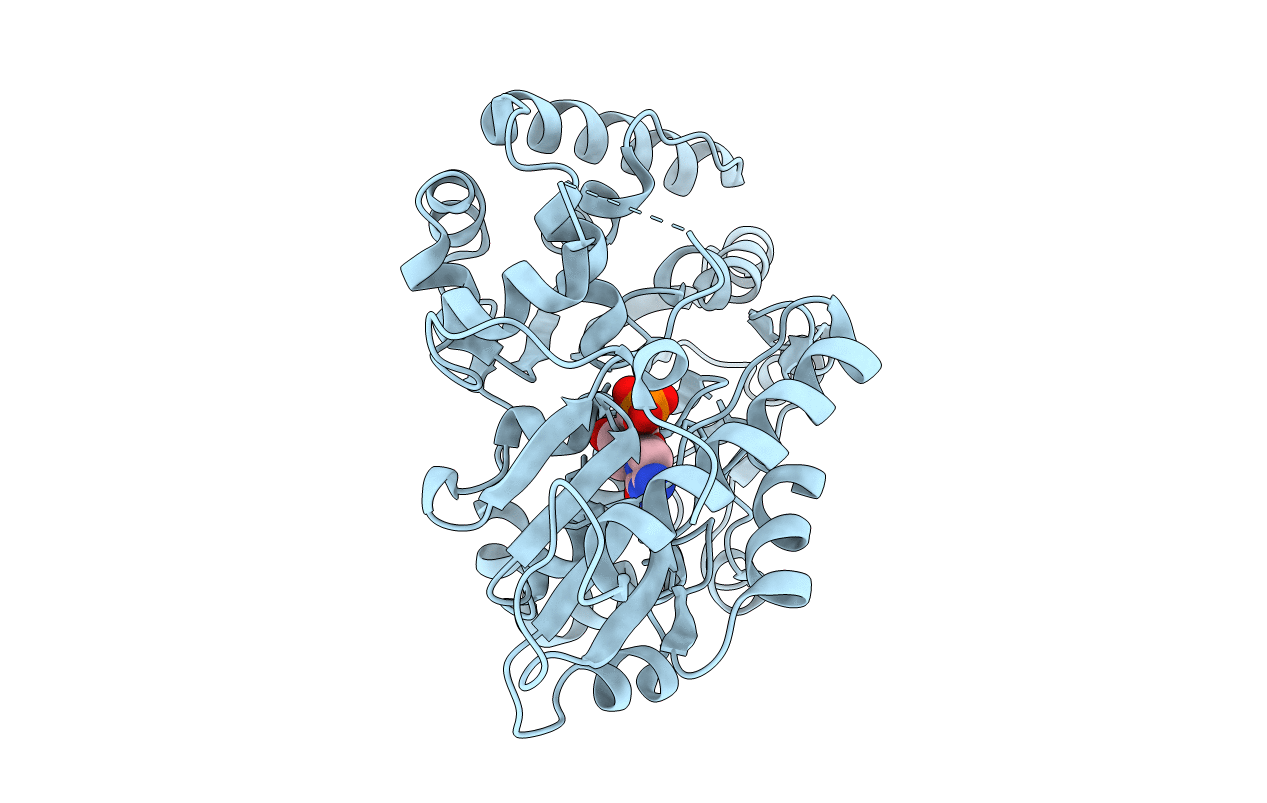
Title:
Crystal structure of mutifunctional sialyltransferase from Pasteurella multocida with CMP bound
Biological Source:
Source Organism(s):
Pasteurella multocida (Taxon ID: 747)
Expression System(s):
Method Details:
Experimental Method:
Resolution:
2.00 Å
R-Value Free:
0.22
R-Value Work:
0.19
R-Value Observed:
0.19
Space Group:
P 1 21 1
