2EWT image
Deposition Date 2005-11-07
Release Date 2006-06-13
Last Version Date 2024-03-13
Entry Detail
PDB ID:
2EWT
Title:
Crystal structure of the DNA-binding domain of BldD
Biological Source:
Source Organism:
Host Organism:
Method Details:
Experimental Method:
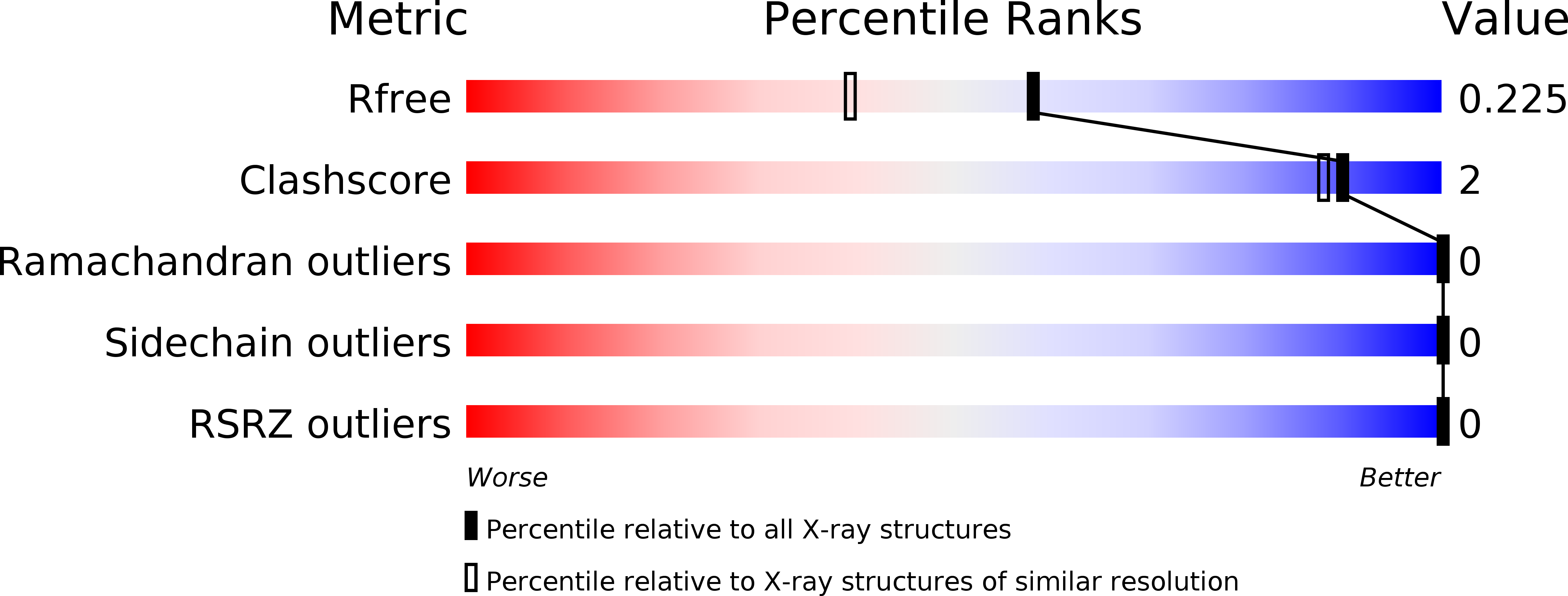
Resolution:
1.81 Å
R-Value Free:
0.22
R-Value Work:
0.18
R-Value Observed:
0.18
Space Group:
C 1 2 1
Macromolecular Entities
Polymer Type:polypeptide(L)
Molecule:putative DNA-binding protein
Chain IDs:A
Chain Length:71
Number of Molecules:1
Biological Source:Streptomyces coelicolor
Ligand Molecules
Primary Citation
Crystal structure of the DNA-binding domain of BldD, a central regulator of aerial mycelium formation in Streptomyces coelicolor A3(2)
Mol.Microbiol. 60 1179 1193 (2006)
PMID: 16689794 DOI: 10.1111/j.1365-2958.2006.05176.x

Abstact

BldD is a central regulator of the developmental process in Streptomyces coelicolor. The 1.8 angstroms resolution structure of the DNA-binding domain of BldD (BldDN) reveals that BldDN forms a compact globular domain composed of four helices (alpha1-alpha4) containing a helix-turn-helix motif (alpha2-alpha3) resembling that of the DNA-binding domain of lambda repressor. The BldDN/DNA complex model led us to design a series of mutants, which revealed the important role of alpha3 and the 'turn' region between alpha2 and alpha3 for DNA recognition. Based on the fact that BldD occupies two operator sites of bldN and whiG and shows significant disparity in the affinity toward the two operator sites when they are disconnected, we propose a model of cooperative binding, which means that the binding of one BldD dimer to the high affinity site facilitates that of the second BldD dimer to the low affinity site. In addition, structural and mutational investigation reveals that the Tyr62Cys mutation, found in the first-identified bldD mutant, can destabilize BldD structure by disrupting the hydrophobic core.

Legend

Protein

Chemical

Disease

Primary Citation of related structures