2EVK image
Deposition Date 2005-10-31
Release Date 2006-04-18
Last Version Date 2023-08-23
Entry Detail
PDB ID:
2EVK
Title:
The Structures of Thiolate- and Carboxylate-Ligated Ferric H93G Myoglobin: Models for Cytochrome P450 and for Oxyanion-Bound Heme Proteins
Biological Source:
Source Organism:
Host Organism:
Method Details:
Experimental Method:
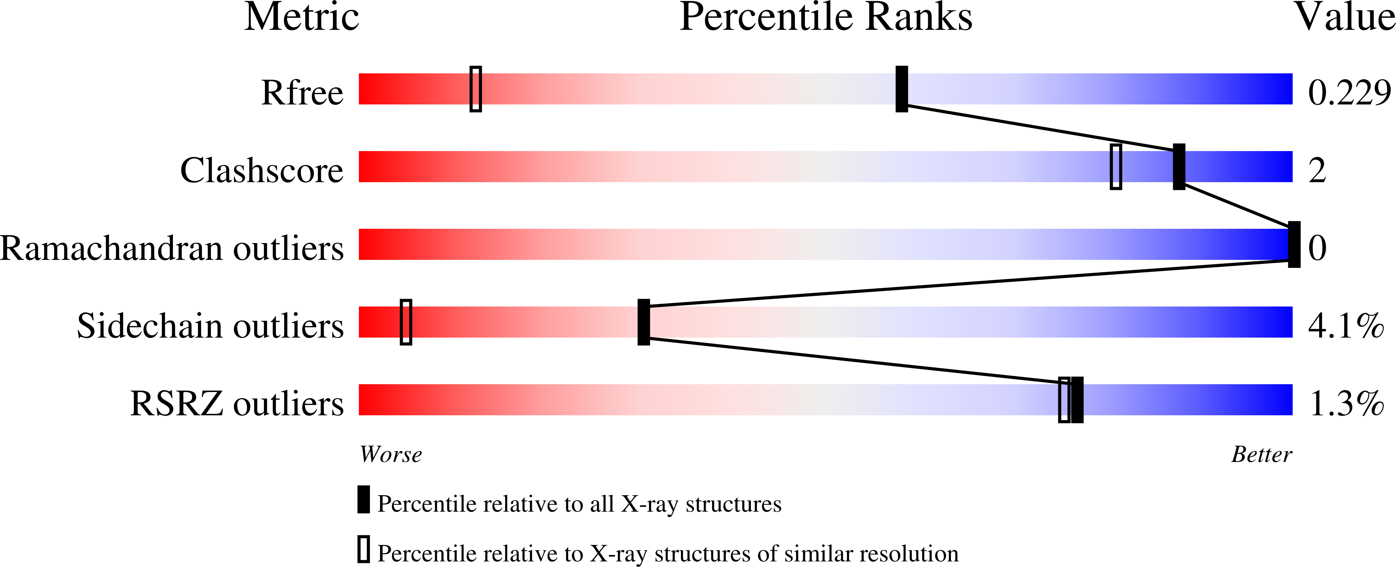
Resolution:
1.40 Å
R-Value Free:
0.26
R-Value Work:
0.20
R-Value Observed:
0.20
Space Group:
P 21 21 21
Macromolecular Entities
Polymer Type:polypeptide(L)
Molecule:Myoglobin
Gene (Uniprot):MB
Mutations:H93G
Chain IDs:A
Chain Length:153
Number of Molecules:1
Biological Source:Physeter catodon
Primary Citation
Structures of thiolate- and carboxylate-ligated ferric H93G myoglobin: models for cytochrome P450 and for oxyanion-bound heme proteins.
Biochemistry 45 3170 3177 (2006)
PMID: 16519512 DOI: 10.1021/bi052171s

Abstact

Crystal structures of the ferric H93G myoglobin (Mb) cavity mutant containing either an anionic proximal thiolate sulfur donor or a carboxylate oxygen donor ligand are reported at 1.7 and 1.4 A resolution, respectively. The crystal structure and magnetic circular dichroism spectra of the H93G Mb beta-mercaptoethanol (BME) thiolate adduct reveal a high-spin, five-coordinate complex. Furthermore, the bound BME appears to have an intramolecular hydrogen bond involving the alcohol proton and the ligated thiolate sulfur, mimicking one of the three proximal N-H...S hydrogen bonds in cytochrome P450. The Fe is displaced from the porphyrin plane by 0.5 A and forms a 2.41 A Fe-S bond. The Fe(3+)-S-C angle is 111 degrees , indicative of a covalent Fe-S bond with sp(3)-hybridized sulfur. Therefore, the H93G Mb.BME complex provides an excellent protein-derived structural model for high-spin ferric P450. In particular, the Fe-S bond in high-spin ferric P450-CAM has essentially the same geometry despite the constraints imposed by covalent linkage of the cysteine to the protein backbone. This suggests that evolution led to the geometric optimization of the proximal Fe-S(cysteinate) bond in P450. The crystal structure and spectral properties of the H93G Mb acetate adduct reveal a high-spin, six-coordinate complex with proximal acetate and distal water axial ligands. The distal His-64 forms a hydrogen bond with the bound water. The Fe-acetate bonding geometry is inconsistent with an electron pair along the Fe-O bond as the Fe-O-C angle is 152 degrees and the Fe is far from the plane of the acetate. Thus, the Fe-O bonding is ionic. The H93G Mb cavity mutant has already been shown to be a versatile model system for the study of ligand binding to heme proteins; this investigation affords the first structural evidence that nonimidazole exogenous ligands bind in the proximal ligation site.

Legend

Protein

Chemical

Disease

Primary Citation of related structures