2EV3 image
Deposition Date 2005-10-30
Release Date 2006-11-07
Last Version Date 2024-03-13
Entry Detail
PDB ID:
2EV3
Keywords:
Title:
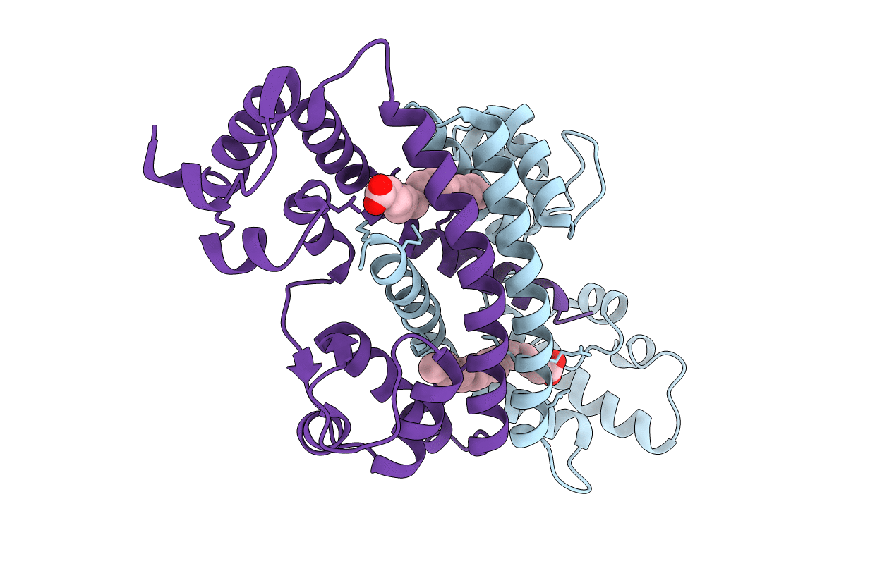
Structure of Rv1264N, the regulatory domain of the mycobacterial adenylyl cylcase Rv1264, at pH 5.3
Biological Source:
Source Organism:
Host Organism:
Method Details:
Experimental Method:
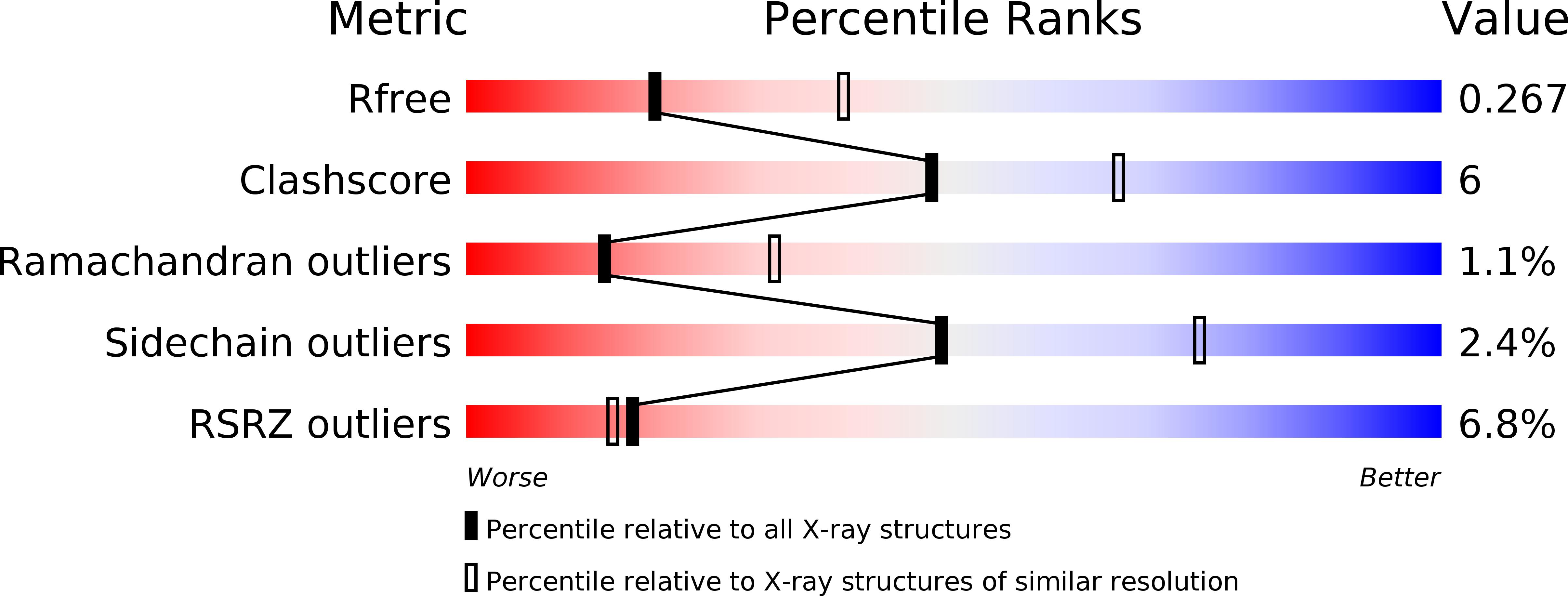
Resolution:
2.68 Å
R-Value Free:
0.26
R-Value Work:
0.22
R-Value Observed:
0.23
Space Group:
C 1 2 1
Macromolecular Entities
Polymer Type:polypeptide(L)
Molecule:Hypothetical protein Rv1264/MT1302
Chain IDs:A, B
Chain Length:222
Number of Molecules:2
Biological Source:Mycobacterium tuberculosis
Ligand Molecules
Primary Citation
The structure of the regulatory domain of the adenylyl cyclase Rv1264 from Mycobacterium tuberculosis with bound oleic acid
J.Mol.Biol. 369 1282 1295 (2007)
PMID: 17482646 DOI: 10.1016/j.jmb.2007.04.013

Abstact

The universal secondary messenger cAMP is produced by adenylyl cyclases (ACs). Most bacterial and all eukaryotic ACs belong to class III of six divergent classes. A class III characteristic is formation of the catalytic pocket at a dimer interface and the presence of additional regulatory domains. Mycobacterium tuberculosis possesses 15 class III ACs, including Rv1264, which is activated at acidic pH due to pH-dependent structural transitions of the Rv1264 dimer. It has been shown by X-ray crystallography that the N-terminal regulatory and C-terminal catalytic domains of Rv1264 interact in completely different ways in the active and inhibited states. Here, we report an in-depth structural and functional analysis of the regulatory domain of Rv1264. The 1.6 A resolution crystal structure shows the protein in a tight, disk-shaped dimer, formed around a helical bundle, and involving a protein chain crossover. To understand pH regulation, we determined structures at acidic and basic pH values and employed structure-based mutagenesis in the holoenzyme to elucidate regulation using an AC activity assay. It has been shown that regulatory and catalytic domains must be linked in a single protein chain. The new studies demonstrate that the length of the linker segment is decisive for regulation. Several amino acids on the surface of the regulatory domain, when exchanged, altered the pH-dependence of AC activity. However, these residues are not conserved amongst a number of related ACs. The closely related mycobacterial Rv2212, but not Rv1264, is strongly activated by the addition of fatty acids. The structure resolved the presence of a deeply embedded fatty acid, characterised as oleic acid by mass spectrometry, which may serve as a hinge. From these data, we conclude that the regulatory domain is a structural scaffold used for distinct regulatory purposes.

Legend

Protein

Chemical

Disease

Primary Citation of related structures