2ETB image
Deposition Date 2005-10-27
Release Date 2006-06-27
Last Version Date 2023-08-23
Entry Detail
PDB ID:
2ETB
Title:
Crystal structure of the ankyrin repeat domain of TRPV2
Biological Source:
Source Organism:
Method Details:
Experimental Method:
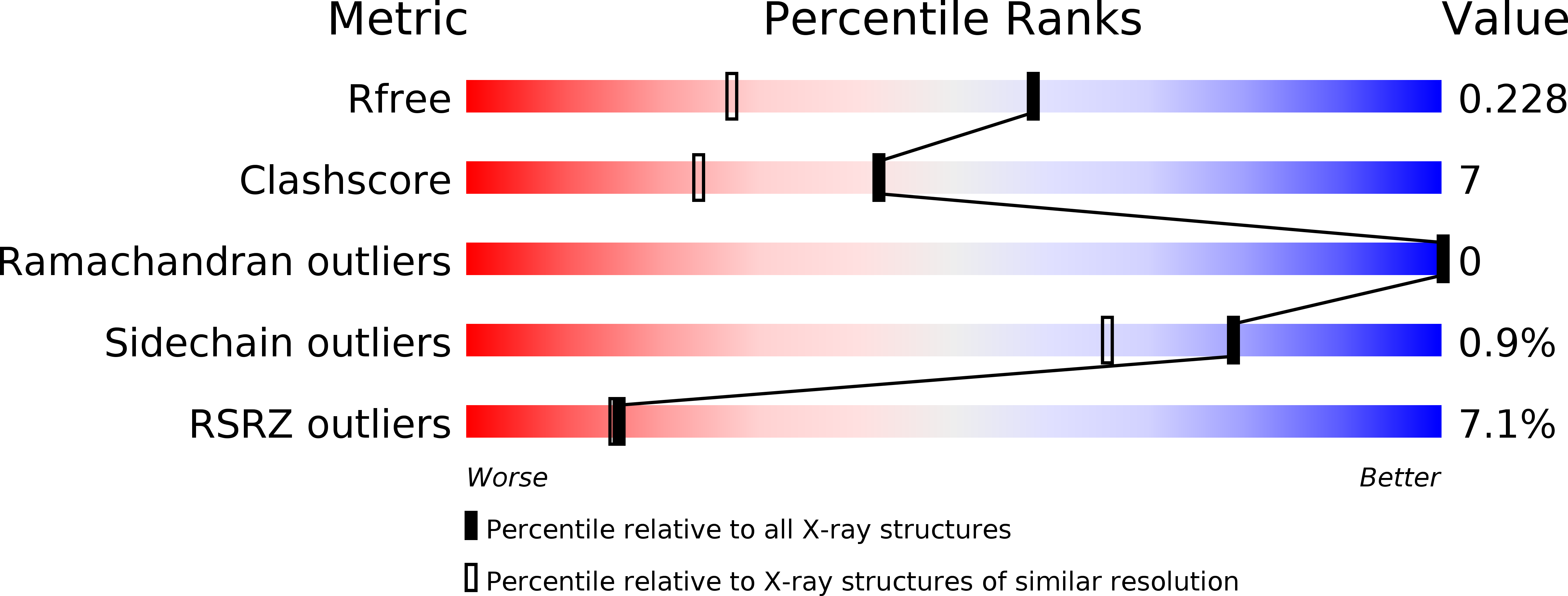
Resolution:
1.65 Å
R-Value Free:
0.22
R-Value Work:
0.18
R-Value Observed:
0.18
Space Group:
P 21 21 21
Macromolecular Entities
Polymer Type:polypeptide(L)
Molecule:Transient receptor potential cation channel subfamily V member 2
Gene (Uniprot):Trpv2
Chain IDs:A
Chain Length:256
Number of Molecules:1
Biological Source:Rattus norvegicus
Ligand Molecules
Primary Citation
Structure of the N-terminal Ankyrin Repeat Domain of the TRPV2 Ion Channel.
J.Biol.Chem. 281 25006 25010 (2006)
PMID: 16809337 DOI: 10.1074/jbc.C600153200

Abstact

The TRPV ion channels mediate responses to many sensory stimuli including heat, low pH, neuropeptides, and chemical ligands. All TRPV subfamily members contain an intracellular N-terminal ankyrin repeat domain (ARD), a prevalent protein interaction motif. The 1.6-A crystal structure of the TRPV2-ARD, with six ankyrin repeats, reveals several atypical structural features. Repeats one through three display unusually long and flexible fingers with a large number of exposed aromatic residues, whereas repeats five and six have unusually long outer helices. Furthermore, a large counterclockwise twist observed in the stacking of repeats four and five breaks the regularity of the domain, altering the shape of surfaces available for interactions with proteins or other cellular ligands. Both solution studies and crystal packing interactions indicate that the TRPV2-ARD does not form homo-oligomers, suggesting that the ARD of TRPV ion channels may be used for interactions with regulatory factors rather than in promoting tetrameric assembly of the ion channels.

Legend

Protein

Chemical

Disease

Primary Citation of related structures