Deposition Date
2005-10-26
Release Date
2005-12-20
Last Version Date
2024-11-20
Entry Detail
PDB ID:
2ESC
Keywords:
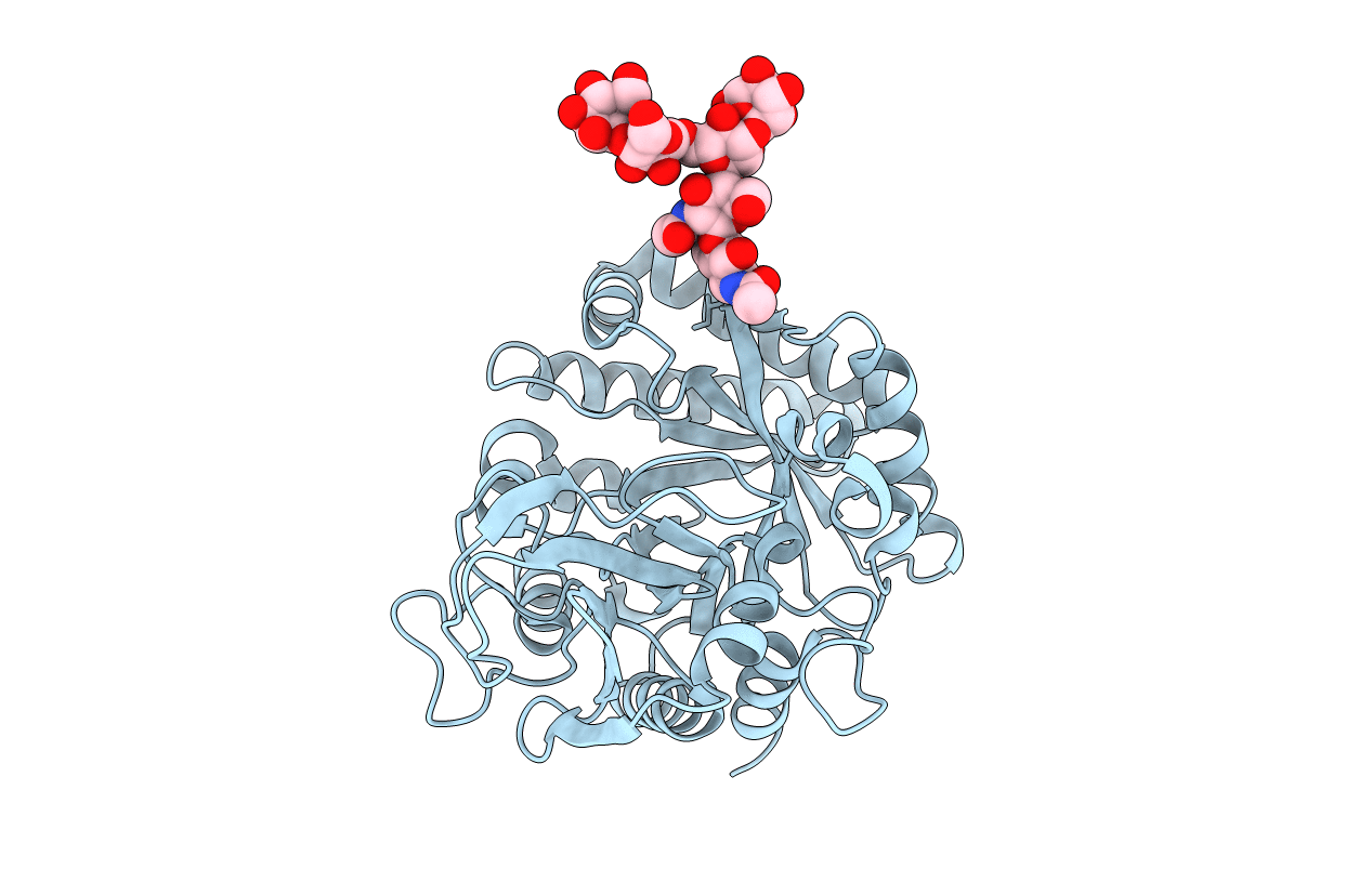
Title:
Crystal structure of a 40 KDa protective signalling protein from Bovine (SPC-40) at 2.1 A resolution
Biological Source:
Source Organism(s):
Bos taurus (Taxon ID: 9913)
Method Details:
Experimental Method:
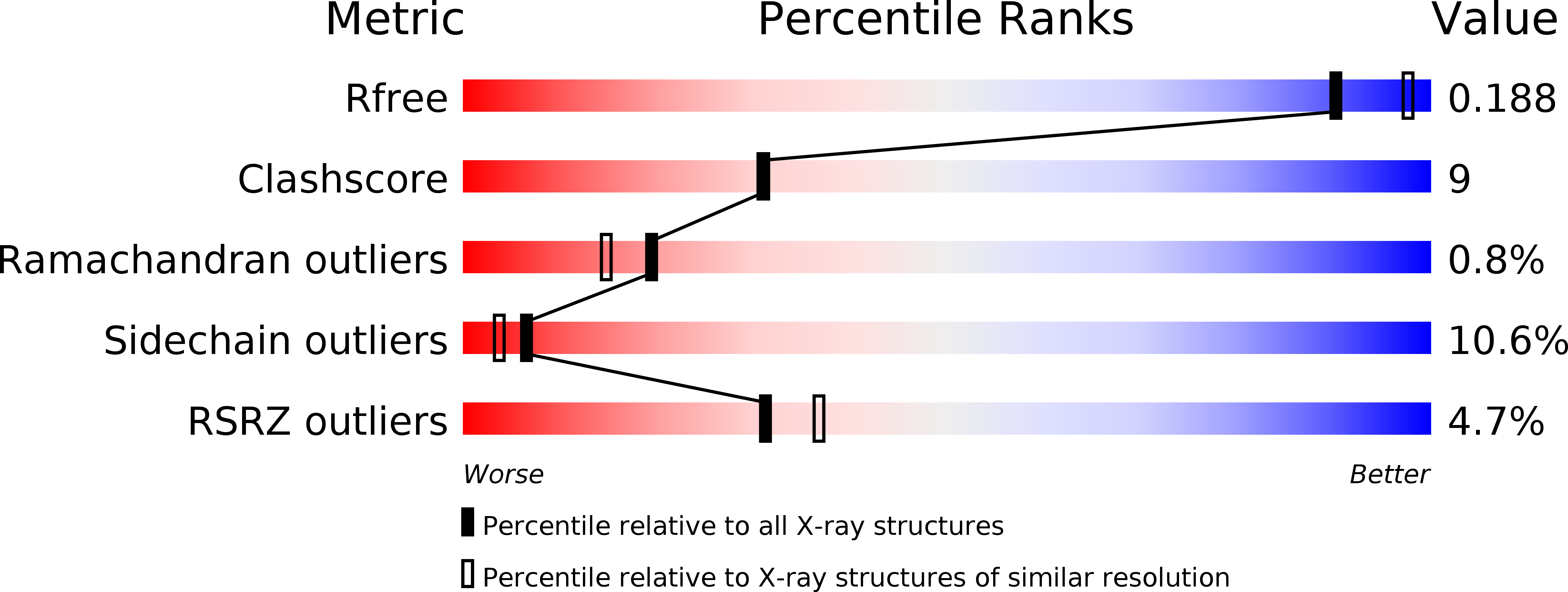
Resolution:
2.10 Å
R-Value Free:
0.22
R-Value Work:
0.17
R-Value Observed:
0.17
Space Group:
P 21 21 21