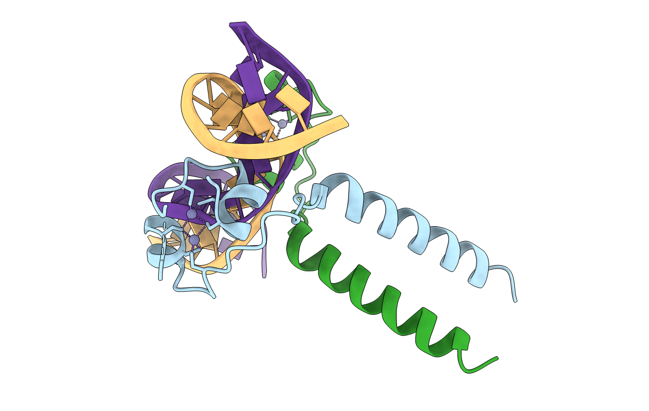
2ERE image
Entry Detail
PDB ID:
2ERE
Title:
Crystal Structure of a Leu3 DNA-binding domain complexed with a 15mer DNA duplex
Biological Source:
Source Organism:
Host Organism:
PDB Version:
Deposition Date:
2005-10-24
Release Date:
2006-04-04
Method Details:
Experimental Method:
Resolution:
3.00 Å
R-Value Free:
0.27
R-Value Work:
0.27
R-Value Observed:
0.27
Space Group:
P 62
Macromolecular Entities
Polymer Type:polypeptide(L)
Description:Regulatory protein LEU3
Chain IDs:C (auth: A), D (auth: B)
Chain Length:72
Number of Molecules:2
Biological Source:Saccharomyces cerevisiae
Ligand Molecules
Primary Citation
Structure of a Leu3-DNA complex: recognition of everted CGG half-sites by a Zn2Cys6 binuclear cluster protein.
Structure 14 725 735 (2006)
PMID: 16615914 DOI: 10.1016/j.str.2005.11.025

Abstact

Gal4 is the prototypical Zn2Cys6 binuclear cluster transcriptional regulator that binds as a homodimer to DNA containing inverted CGG half-sites. Leu3, a member of this protein family, binds to everted (opposite polarity to inverted) CGG half-sites, and an H50C mutation within the Leu3 Zn2Cys6 binuclear motif abolishes its transcriptional repression function without impairing DNA binding. We report the X-ray crystal structures of DNA complexes with Leu3 and Leu3(H50C) and solution DNA binding studies of selected Leu3 mutant proteins. These studies reveal the molecular details of everted CGG half-site recognition, and suggest a role for the H50C mutation in transcriptional repression. Comparison with the Gal4-DNA complex shows an unexpected conservation in the DNA recognition mode of inverted and everted CGG half-sites, and points to a critical function of a linker region between the Zn2Cys6 binuclear cluster and dimerization regions in DNA binding specificity. Broader implications of these findings are discussed.

Legend

Protein

Chemical

Disease

Primary Citation of related structures