Deposition Date
2007-03-28
Release Date
2008-04-01
Last Version Date
2023-10-25
Entry Detail
PDB ID:
2EMS
Keywords:
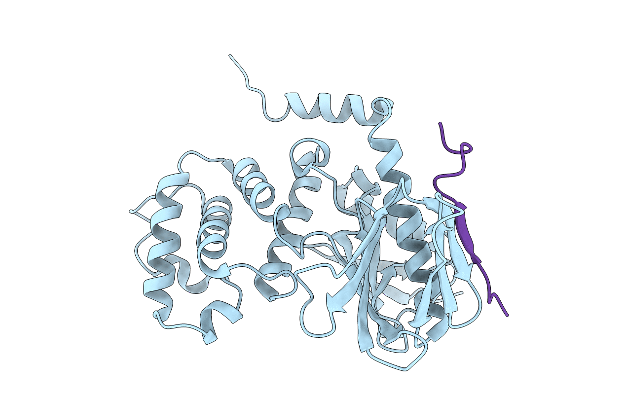
Title:
Crystal Structure Analysis of the radixin FERM domain complexed with adhesion molecule CD43
Biological Source:
Source Organism(s):
Mus musculus (Taxon ID: 10090)
Expression System(s):
Method Details:
Experimental Method:
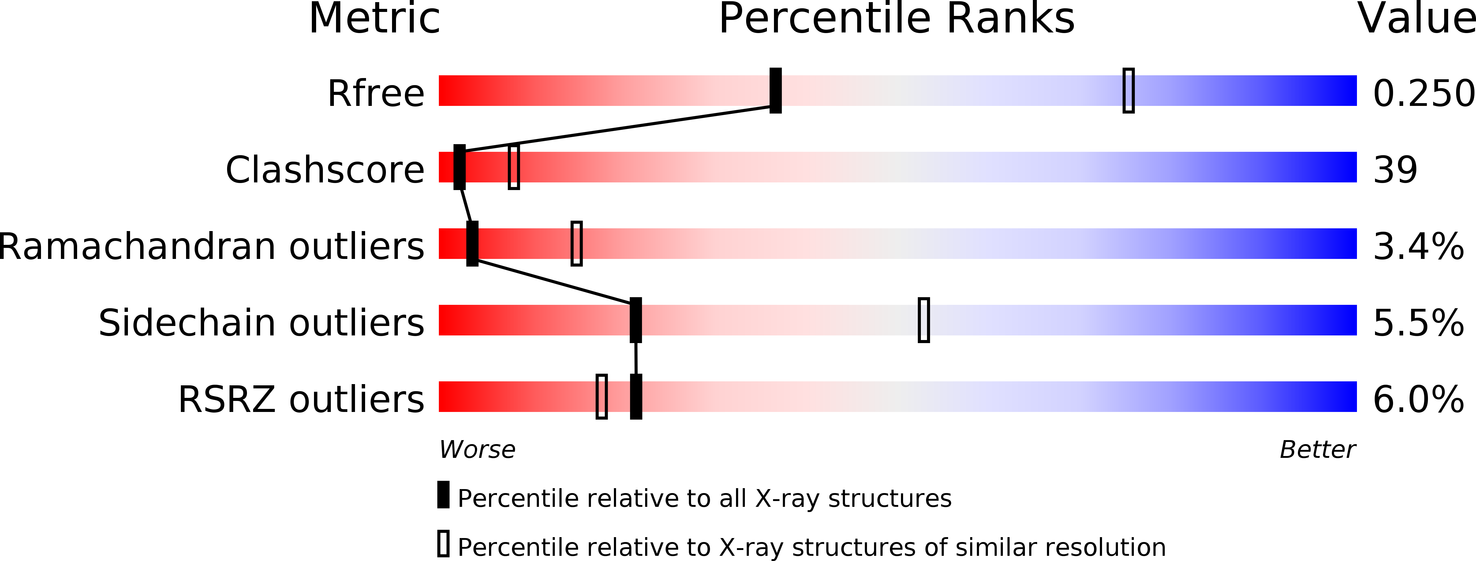
Resolution:
2.90 Å
R-Value Free:
0.25
R-Value Work:
0.23
R-Value Observed:
0.23
Space Group:
P 43 2 2