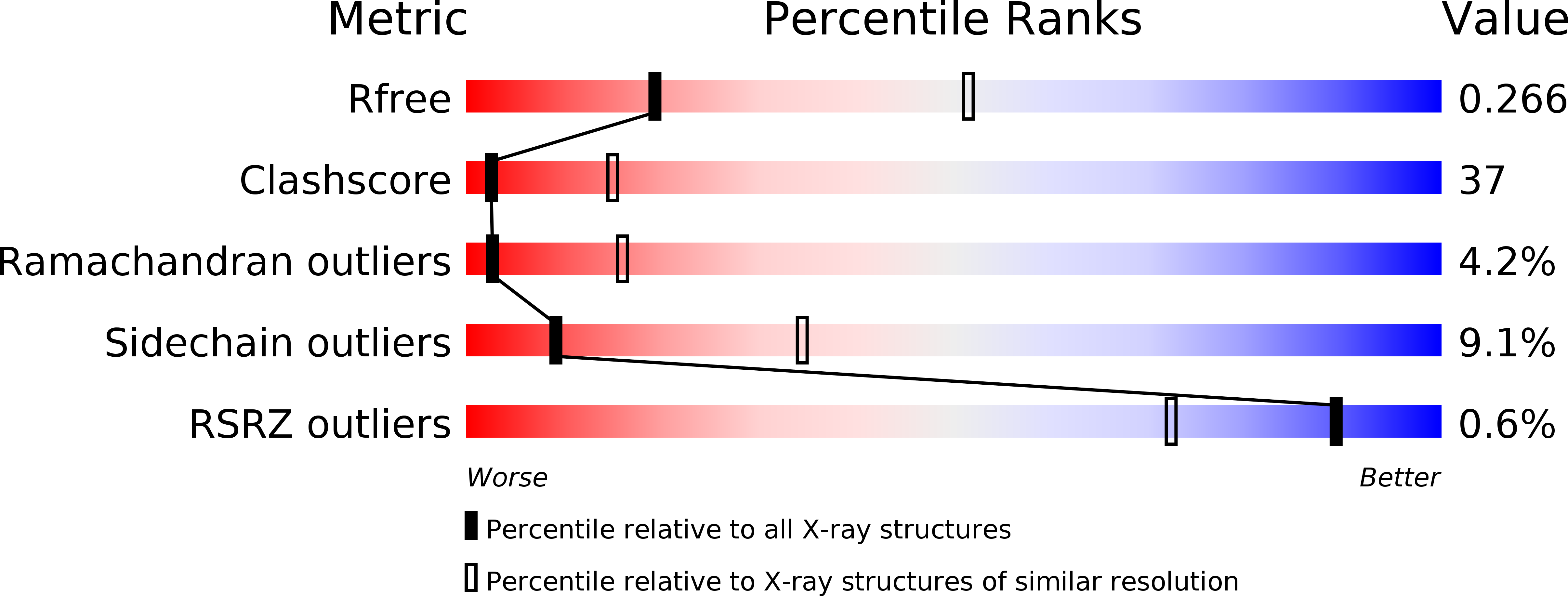
2EHO image
Deposition Date 2007-03-07
Release Date 2007-06-19
Last Version Date 2024-10-30
Entry Detail
PDB ID:
2EHO
Keywords:
Title:
Crystal structure of human GINS complex
Biological Source:
Source Organism:
Homo sapiens (Taxon ID: 9606)
Method Details:
Experimental Method:
Resolution:
3.00 Å
R-Value Free:
0.27
R-Value Work:
0.22
R-Value Observed:
0.22
Space Group:
C 1 2 1
Macromolecular Entities
Structures with similar UniProt ID
Protein Blast
Polymer Type:polypeptide(L)
Molecule:GINS complex subunit 4
Gene (Uniprot):GINS4
Chain IDs:A, E, I
Chain Length:203
Number of Molecules:3
Biological Source:Homo sapiens
Structures with similar UniProt ID
Protein Blast
Polymer Type:polypeptide(L)
Molecule:DNA replication complex GINS protein PSF1
Gene (Uniprot):GINS1
Chain IDs:B, F, J
Chain Length:152
Number of Molecules:3
Biological Source:Homo sapiens
Structures with similar UniProt ID
Protein Blast
Polymer Type:polypeptide(L)
Molecule:DNA replication complex GINS protein PSF2
Gene (Uniprot):GINS2
Chain IDs:C, G, K
Chain Length:186
Number of Molecules:3
Biological Source:Homo sapiens
Structures with similar UniProt ID
Protein Blast
Polymer Type:polypeptide(L)
Molecule:GINS complex subunit 3
Gene (Uniprot):GINS3
Chain IDs:D, H, L
Chain Length:216
Number of Molecules:3
Biological Source:Homo sapiens
Modified Residue
Compound ID Chain ID Parent Comp ID Details 2D Image
MSE A MET SELENOMETHIONINE
Ligand Molecules
Primary Citation
Crystal structure of the human GINS complex
Genes Dev. 21 1316 1321 (2007)
PMID: 17545466 DOI: 10.1101/gad.1548107

Abstact

The GINS complex mediates the assembly of the MCM2-7 (minichromosome maintenance) complex with proteins in a replisome progression complex. The eukaryotic GINS complex is composed of Sld5, Psf1, Psf2, and Psf3, which must be assembled for cell proliferation. We determined the crystal structure of the human GINS complex: GINS forms an elliptical shape with a small central channel. The structures of Sld5 and Psf2 resemble those of Psf1 and Psf3, respectively. In addition, the N-terminal and C-terminal domains of Sld5/Psf1 are permuted in Psf2/Psf3, which suggests that the four proteins have evolved from a common ancestor. Using a structure-based mutational analysis, we identified the functionally critical surface regions of the GINS complex.

Legend

Protein

Chemical

Disease

Primary Citation of related structures