Deposition Date
2007-02-28
Release Date
2008-03-11
Last Version Date
2023-10-25
Entry Detail
PDB ID:
2EGD
Keywords:
Title:
Crystal structure of human S100A13 in the Ca2+-bound state
Biological Source:
Source Organism(s):
Homo sapiens (Taxon ID: 9606)
Expression System(s):
Method Details:
Experimental Method:
Resolution:
1.80 Å
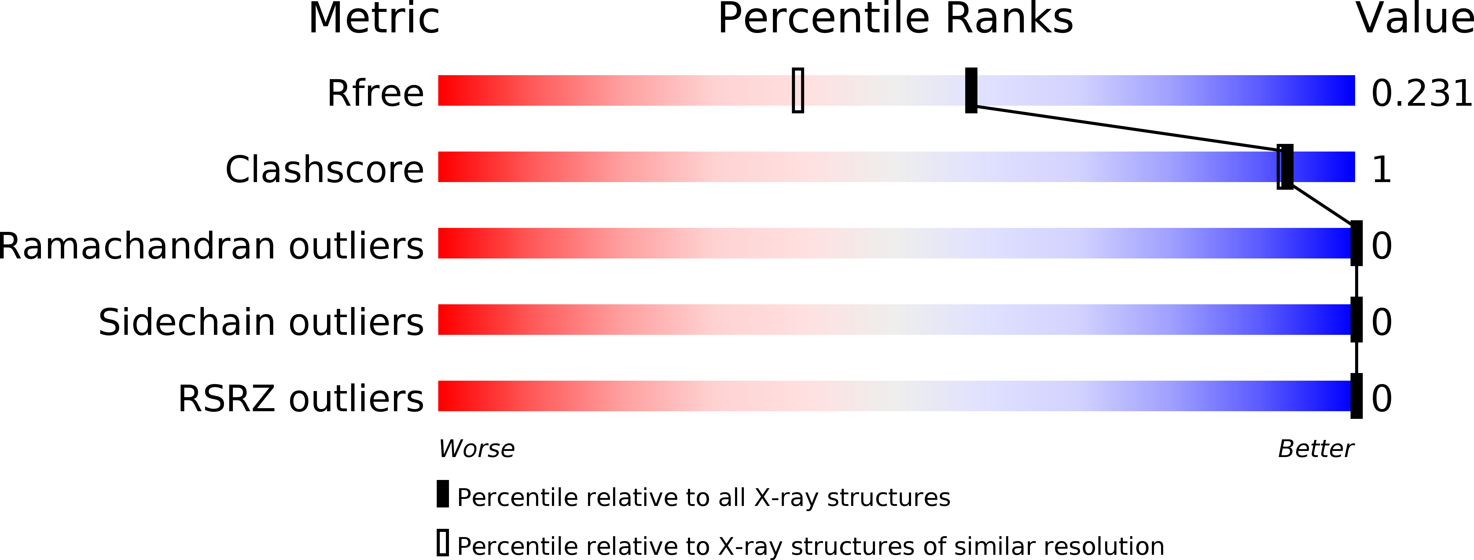
R-Value Free:
0.23
R-Value Work:
0.19
R-Value Observed:
0.19
Space Group:
P 21 21 21