
Deposition Date
2007-02-26
Release Date
2007-08-28
Last Version Date
2024-03-13
Entry Detail
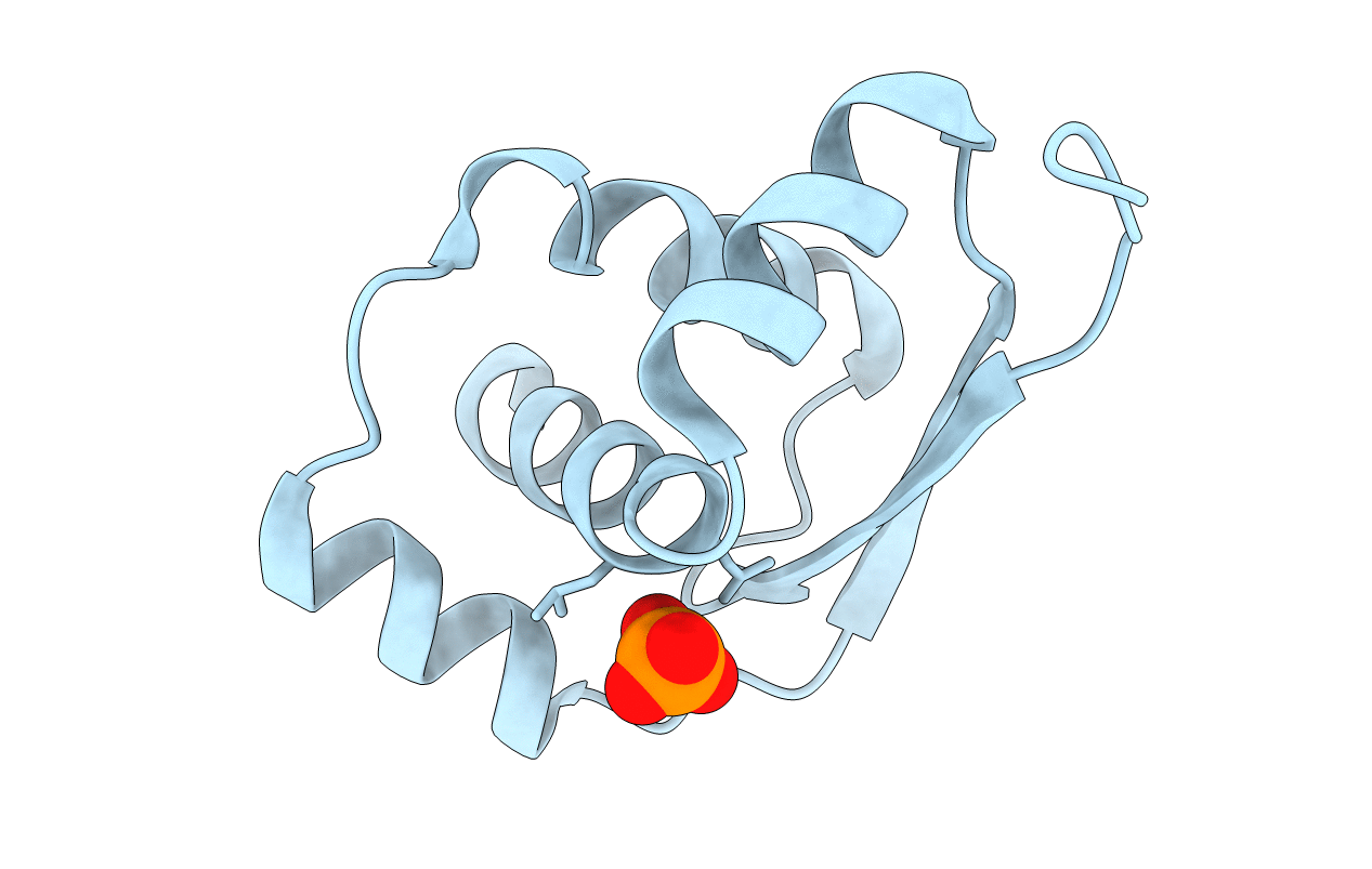
PDB ID:
2EFV
Keywords:
Title:
Crystal Structure of a Hypothetical Protein(MJ0366) from Methanocaldococcus jannaschii
Biological Source:
Source Organism(s):
Methanocaldococcus jannaschii DSM 2661 (Taxon ID: 243232)
Expression System(s):
Method Details:
Experimental Method:
Resolution:
1.90 Å
R-Value Free:
0.23
R-Value Work:
0.21
R-Value Observed:
0.21
Space Group:
P 43 21 2
