
Deposition Date
2007-02-23
Release Date
2007-05-29
Last Version Date
2024-11-20
Entry Detail
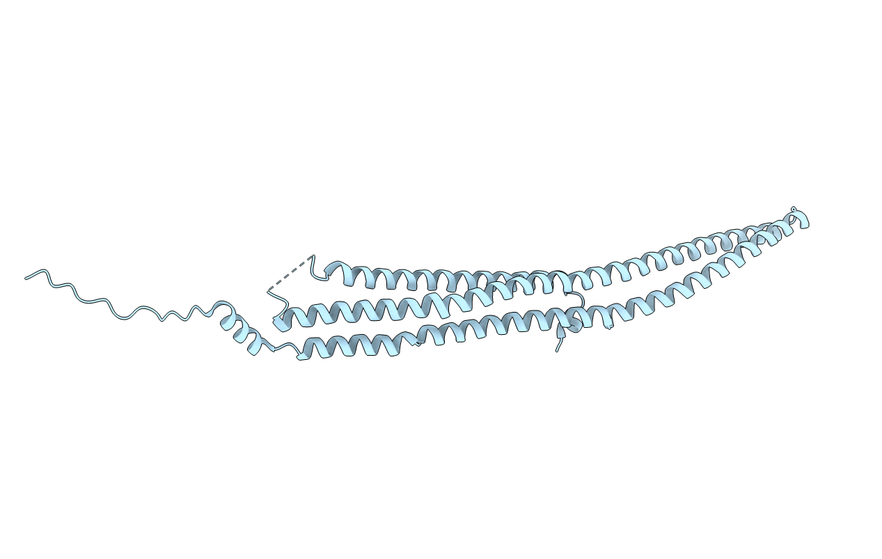
PDB ID:
2EFL
Keywords:
Title:
Crystal structure of the EFC domain of formin-binding protein 17
Biological Source:
Source Organism(s):
Homo sapiens (Taxon ID: 9606)
Method Details:
Experimental Method:
Resolution:
2.61 Å
R-Value Free:
0.26
R-Value Work:
0.21
R-Value Observed:
0.21
Space Group:
C 1 2 1
