
Deposition Date
2007-02-18
Release Date
2007-11-13
Last Version Date
2023-10-25
Entry Detail
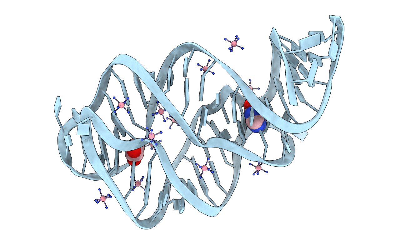
Method Details:
Experimental Method:
Resolution:
1.75 Å
R-Value Free:
0.24
R-Value Work:
0.20
Space Group:
C 1 2 1
