Deposition Date
1998-10-27
Release Date
1999-06-15
Last Version Date
2025-03-26
Entry Detail
PDB ID:
2ECP
Keywords:
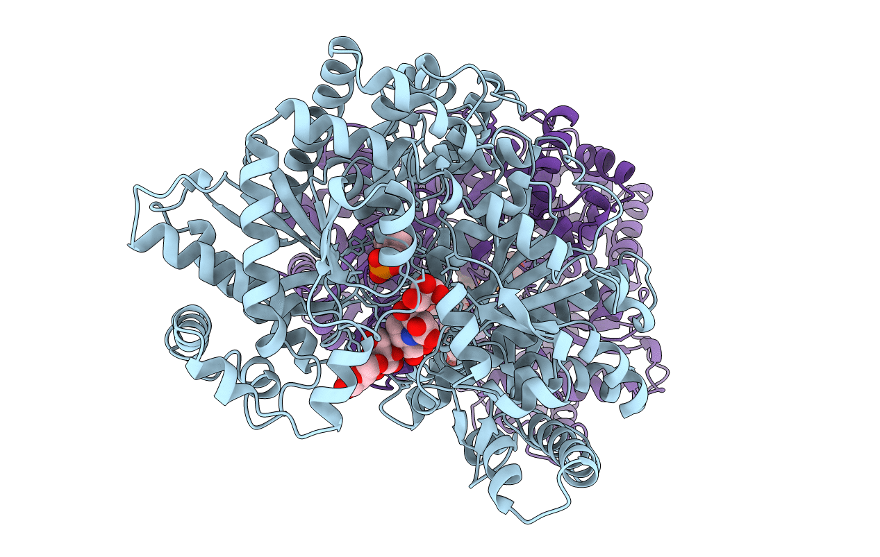
Title:
THE CRYSTAL STRUCTURE OF THE E. COLI MALTODEXTRIN PHOSPHORYLASE COMPLEX
Biological Source:
Source Organism(s):
Escherichia coli (Taxon ID: 562)
Expression System(s):
Method Details:
Experimental Method:
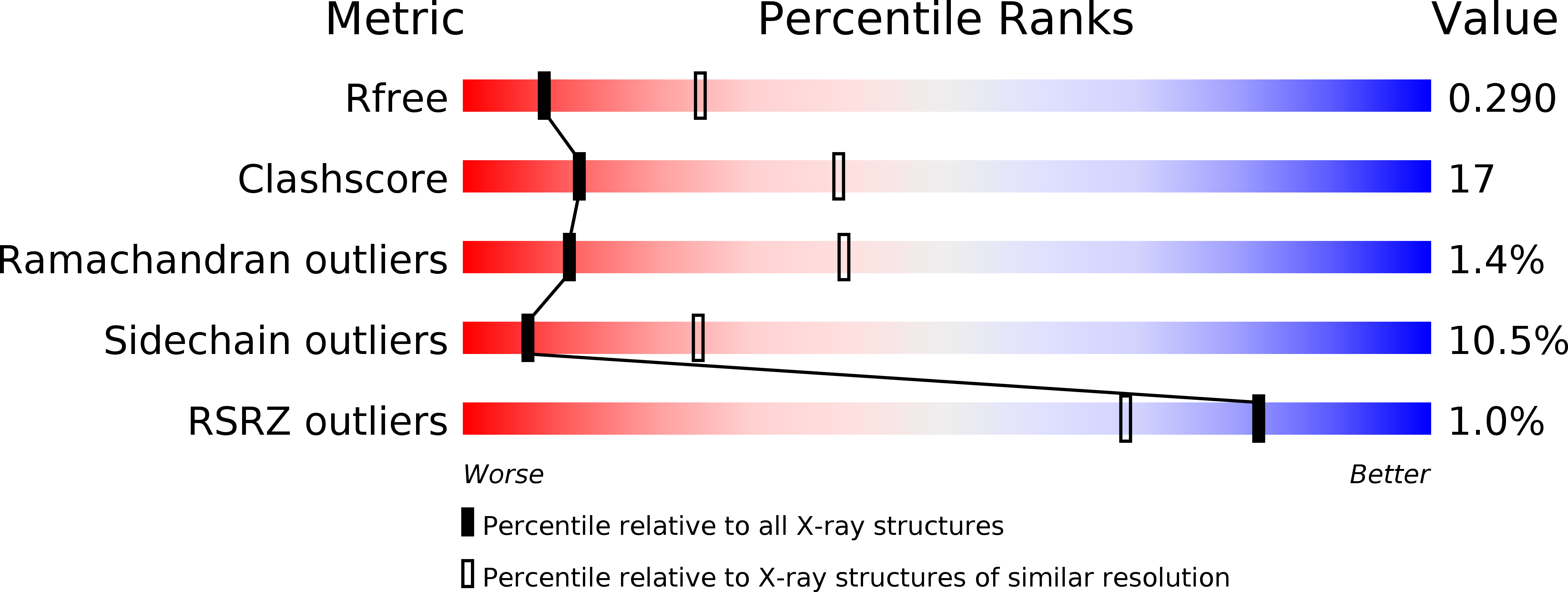
Resolution:
2.95 Å
R-Value Free:
0.29
R-Value Work:
0.24
Space Group:
P 21 21 21