2EB9 image
Deposition Date 2007-02-07
Release Date 2007-07-03
Last Version Date 2023-10-25
Entry Detail
PDB ID:
2EB9
Title:
Crystal Structure of Cu(II)(Sal-Leu)/apo-Myoglobin
Biological Source:
Source Organism:
Host Organism:
Method Details:
Experimental Method:
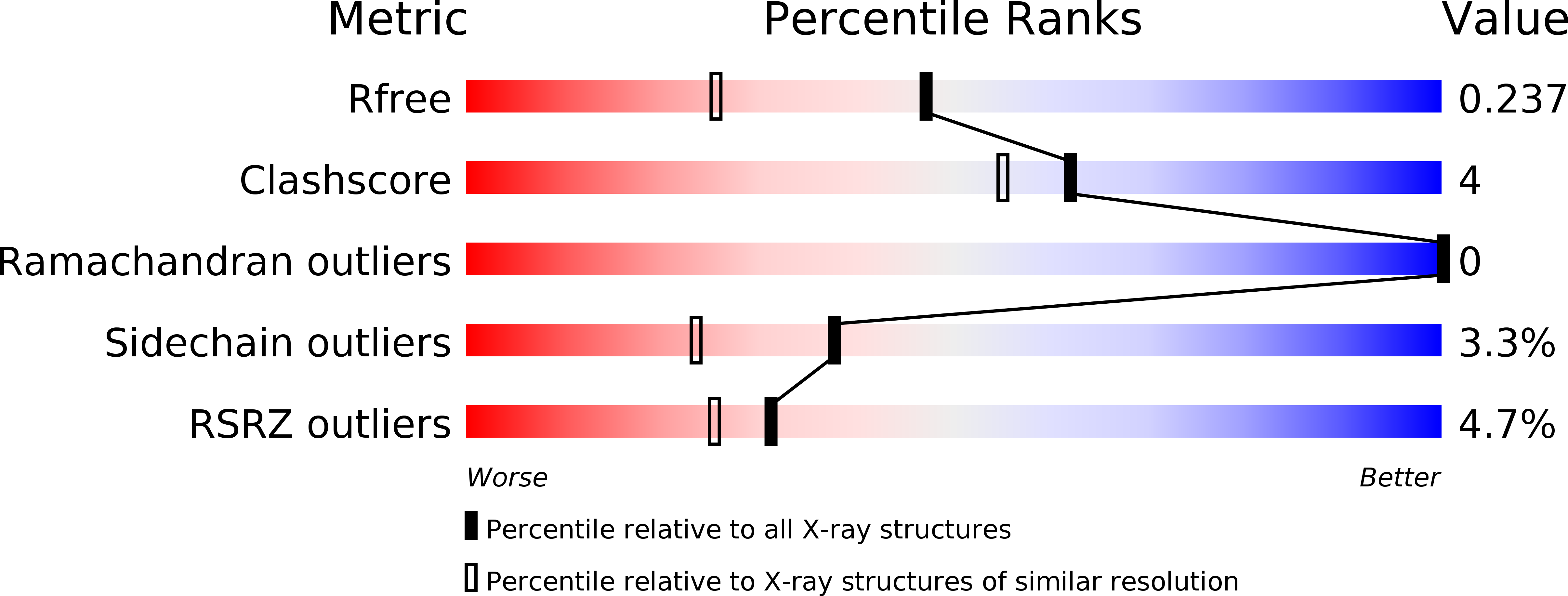
Resolution:
1.80 Å
R-Value Free:
0.23
R-Value Work:
0.18
R-Value Observed:
0.19
Space Group:
P 21 21 21
Macromolecular Entities
Polymer Type:polypeptide(L)
Molecule:Myoglobin
Gene (Uniprot):MB
Chain IDs:A
Chain Length:154
Number of Molecules:1
Biological Source:Physeter catodon
Primary Citation
Design and Structure Analysis of Artificial Metalloproteins: Selective Coordination of His64 to Copper Complexes with Square-Planar Structure in the apo-Myoglobin Scaffold
Inorg.Chem. 46 5137 5139 (2007)
PMID: 17523632 DOI: 10.1021/ic070289m

Abstact

apo-Myoglobin (apo-Mb) was reconstituted with three copper complexes: CuII(Sal-Phe) (1; Sal-Phe = N-salicylidene-L-phenylalanato), CuII(Sal-Leu) (2; Sal-Leu = N-salicylidene-L-leucinato), and CuII(Sal-Ala) (3; Sal-Ala = N-salicylidene-L-alanato). The crystal structures of 1.apo-Mb (1.65 Angstrom resolution) and 2.apo-Mb (1.8 Angstrom resolution) show that the coordination geometry around the CuII atom in apo-Mb is distorted square-planar with tridentate Sal-X and a Nepsilon atom of His64 in the apo-Mb cavity and the plane of these copper complexes is perpendicular to that of heme. These results suggest that the apo-Mb cavity can hold metal complexes with various coordination geometries.

Legend

Protein

Chemical

Disease

Primary Citation of related structures