2E7Q image
Deposition Date 2007-01-12
Release Date 2007-06-19
Last Version Date 2024-10-23
Entry Detail
PDB ID:
2E7Q
Title:
Crystal structure of basic winged bean lectin in complex with b blood group trisaccharide
Biological Source:
Source Organism:
Method Details:
Experimental Method:
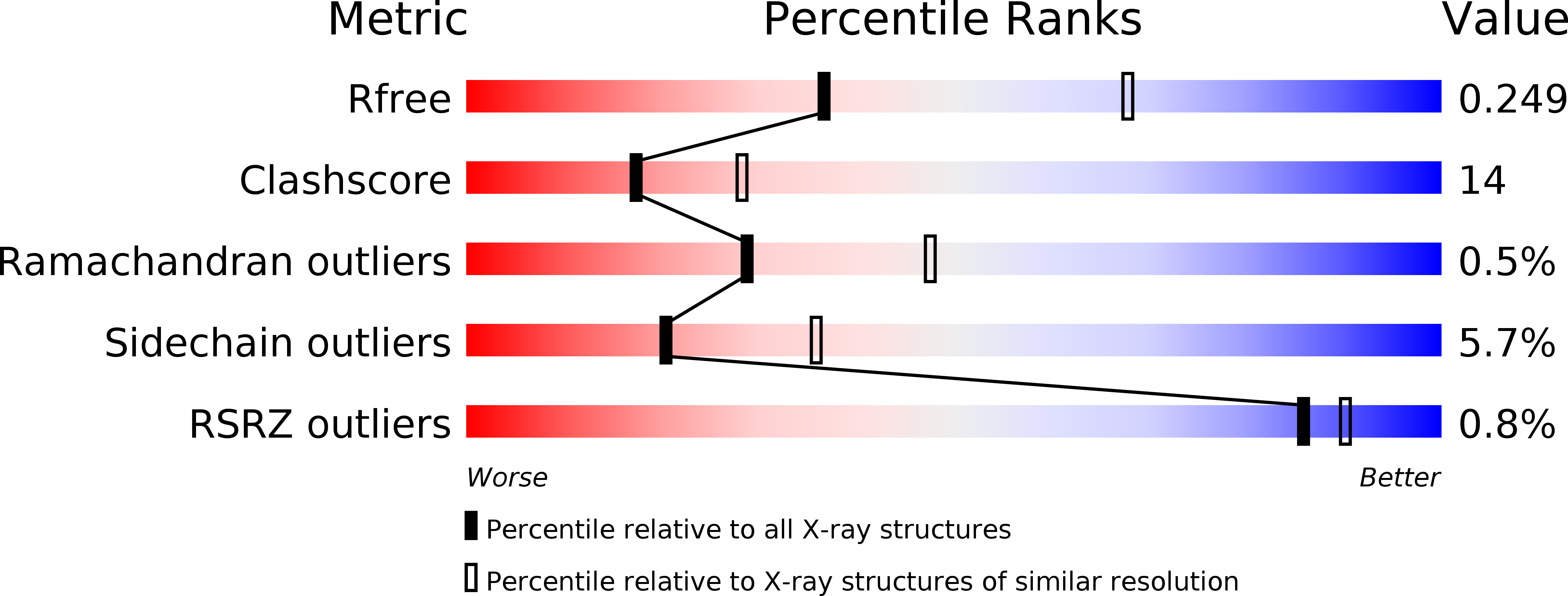
Resolution:
2.75 Å
R-Value Free:
0.25
R-Value Work:
0.19
R-Value Observed:
0.19
Space Group:
P 21 21 2
Macromolecular Entities
Polymer Type:polypeptide(L)
Molecule:Basic agglutinin
Gene (Uniprot):WBAI
Chain IDs:A, B, C, D
Chain Length:237
Number of Molecules:4
Biological Source:Psophocarpus tetragonolobus
Modified Residue
Compound ID Chain ID Parent Comp ID Details 2D Image
ASN A ASN GLYCOSYLATION SITE
Primary Citation
Generation of blood group specificity: New insights from structural studies on the complexes of A- and B-reactive saccharides with basic winged bean agglutinin
Proteins 68 762 769 (2007)
PMID: 17510954 DOI: 10.1002/prot.21428

Abstact

Basic winged bean agglutinin binds A-blood group substance with higher affinity and B-blood group substance with lesser affinity. It does not bind the O substance. The crystal structures of the lectin, complexed with A-reactive and B-reactive di and tri saccharides, have been determined. In addition, the complexes of the lectin with fucosylated A-trisaccharides and B-trisaccharides and with a variant of the A-trisaccharide have been modeled. These structures and models provide valuable insights into the structural basis of blood group specificities. All the four carbohydrate binding loops of the lectin contribute to the primary combining site while the loop of variable length contributes to the secondary binding site. In a significant advance to the current understanding, the interactions at the secondary binding site also contribute substantially, albeit in a subtle manner, to determine the blood group specificity. Compared with the interactions of the B-trisaccharide with the lectin, the third sugar residue of the A-reactive trisacharide forms an additional hydrogen bond with a lysine residue in the variable loop. In the former, the formation of such a hydrogen bond is prevented by a shift in the orientation of third sugar resulting from an internal hydrogen bond in it. The formation of this bond is also facilitated by an interaction dependent change in the rotamer conformation of the lysyl residue of the variable loop. Thus, the difference in the interactions at the secondary site is generated by coordinated movements in the ligand as well as the protein. A comparison of the crystal structure and the model of the complex involving the variant of the A-trisaccharide results in the delineation of the relative contributions of the interactions at the primary and the secondary sites in determining blood group specificity.

Legend

Protein

Chemical

Disease

Primary Citation of related structures