Deposition Date
2006-12-18
Release Date
2007-12-18
Last Version Date
2024-03-13
Entry Detail
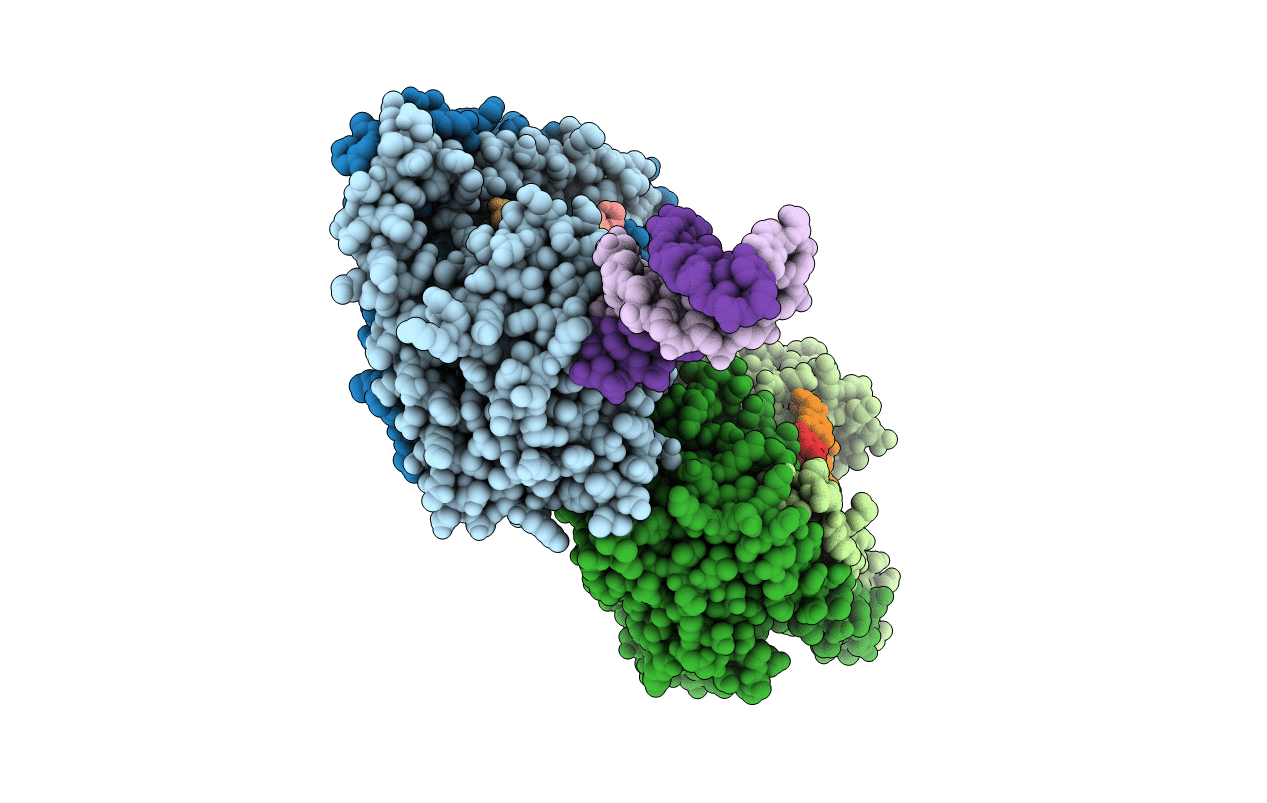
PDB ID:
2E52
Keywords:
Title:
Crystal structural analysis of HindIII restriction endonuclease in complex with cognate DNA at 2.0 angstrom resolution
Biological Source:
Source Organism(s):
Haemophilus influenzae (Taxon ID: 71421)
Expression System(s):
Method Details:
Experimental Method:
Resolution:
2.00 Å
R-Value Free:
0.21
R-Value Work:
0.17
R-Value Observed:
0.17
Space Group:
P 1 21 1