2E4W image
Deposition Date 2006-12-17
Release Date 2007-02-27
Last Version Date 2024-10-23
Entry Detail
PDB ID:
2E4W
Title:
Crystal structure of the extracellular region of the group II metabotropic glutamate receptor complexed with 1S,3S-ACPD
Biological Source:
Source Organism:
Host Organism:
Method Details:
Experimental Method:
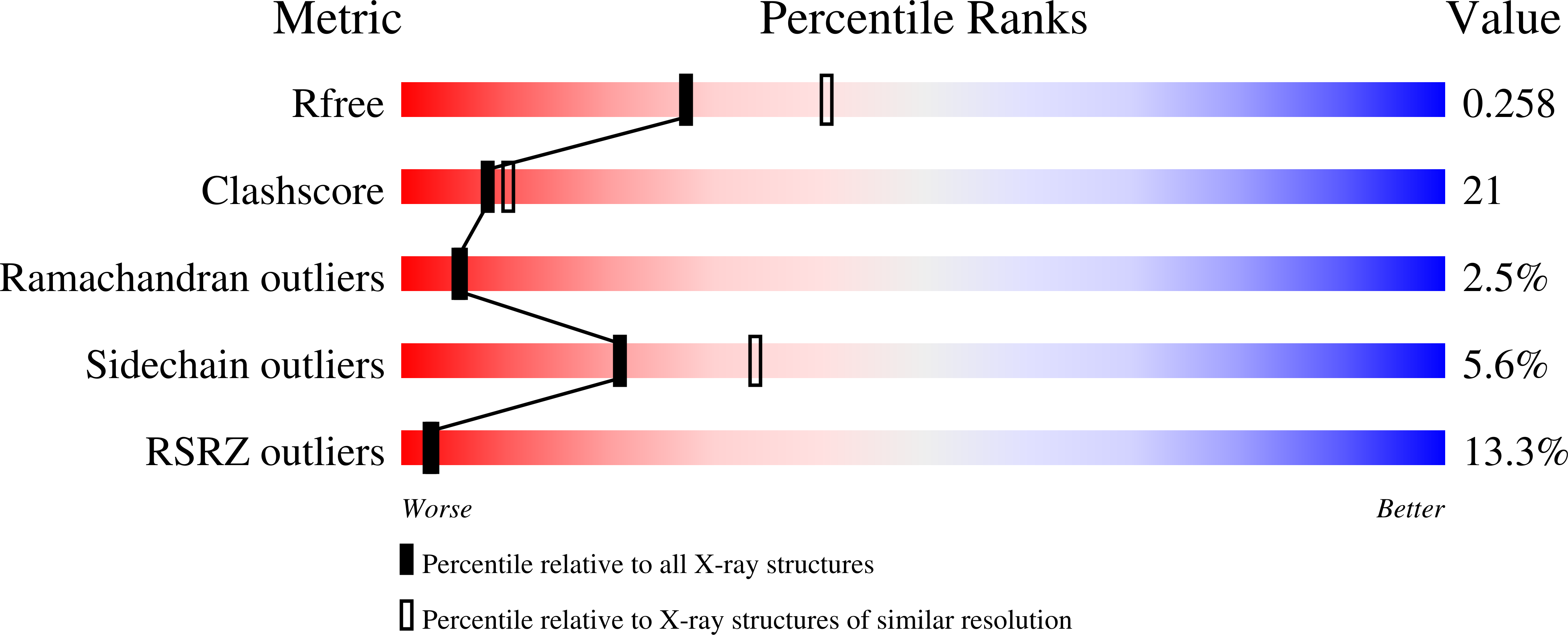
Resolution:
2.40 Å
R-Value Free:
0.26
R-Value Work:
0.23
R-Value Observed:
0.23
Space Group:
P 1 21 1
Macromolecular Entities
Polymer Type:polypeptide(L)
Molecule:Metabotropic glutamate receptor 3
Gene (Uniprot):Grm3
Mutations:N414Q, N439Q
Chain IDs:A, B
Chain Length:555
Number of Molecules:2
Biological Source:Rattus norvegicus
Modified Residue
Compound ID Chain ID Parent Comp ID Details 2D Image
ASN A ASN GLYCOSYLATION SITE
Primary Citation
Structures of the extracellular regions of the group II/III metabotropic glutamate receptors
Proc.Natl.Acad.Sci.Usa 104 3759 3764 (2007)
PMID: 17360426 DOI: 10.1073/pnas.0611577104

Abstact

Metabotropic glutamate receptors play major roles in the activation of excitatory synapses in the central nerve system. We determined the crystal structure of the entire extracellular region of the group II receptor and that of the ligand-binding region of the group III receptor. A comparison among groups I, II, and III provides the structural basis that could account for the discrimination of group-specific agonists. Furthermore, the structure of group II includes the cysteine-rich domain, which is tightly linked to the ligand-binding domain by a disulfide bridge, suggesting a potential role in transmitting a ligand-induced conformational change into the downstream transmembrane region. The structure also reveals the lateral interaction between the two cysteine-rich domains, which could stimulate clustering of the dimeric receptors on the cell surface. We propose a general activation mechanism of the dimeric receptor coupled with both ligand-binding and interprotomer rearrangements.

Legend

Protein

Chemical

Disease

Primary Citation of related structures