Deposition Date
2006-12-01
Release Date
2007-03-20
Last Version Date
2023-10-25
Entry Detail
PDB ID:
2E3Z
Keywords:
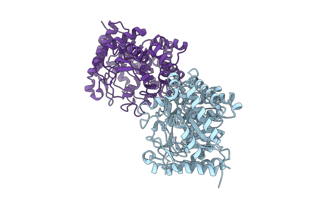
Title:
Crystal structure of intracellular family 1 beta-glucosidase BGL1A from the basidiomycete Phanerochaete chrysosporium in substrate-free form
Biological Source:
Source Organism(s):
Phanerochaete chrysosporium (Taxon ID: 5306)
Expression System(s):
Method Details:
Experimental Method:
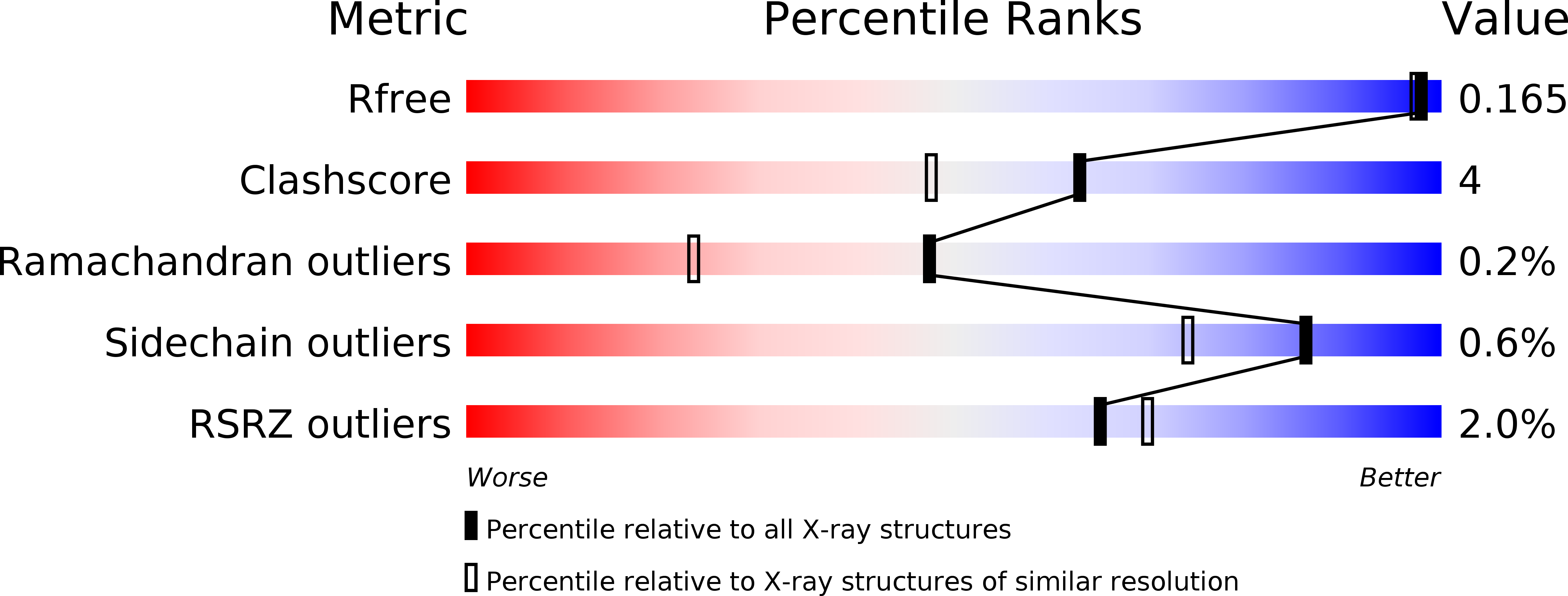
Resolution:
1.50 Å
R-Value Free:
0.18
R-Value Work:
0.16
R-Value Observed:
0.16
Space Group:
P 21 21 21