2E3J image
Deposition Date 2006-11-27
Release Date 2007-12-04
Last Version Date 2023-10-25
Entry Detail
PDB ID:
2E3J
Keywords:
Title:
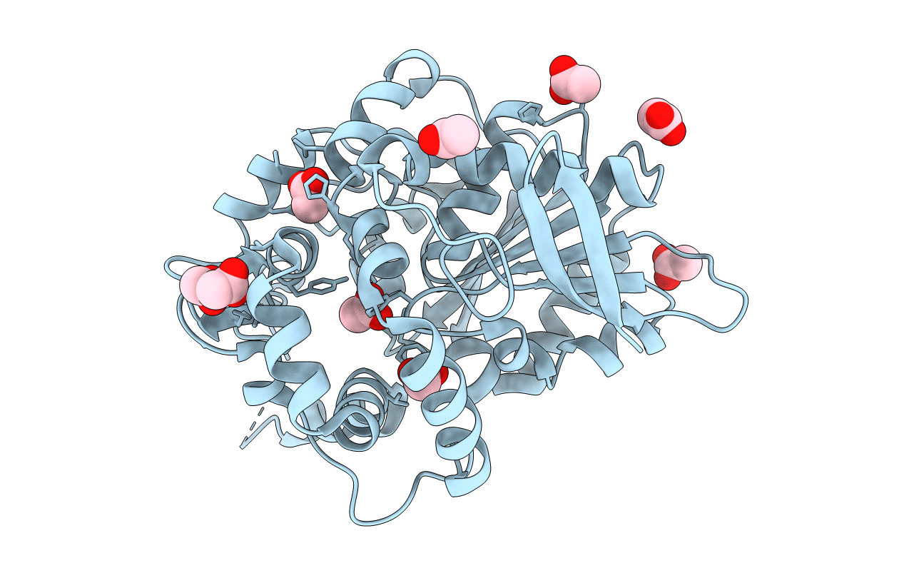
The crystal structure of epoxide hydrolase B (Rv1938) from mycobacterium tuberculosis at 2.1 angstrom
Biological Source:
Source Organism:
Host Organism:
Method Details:
Experimental Method:
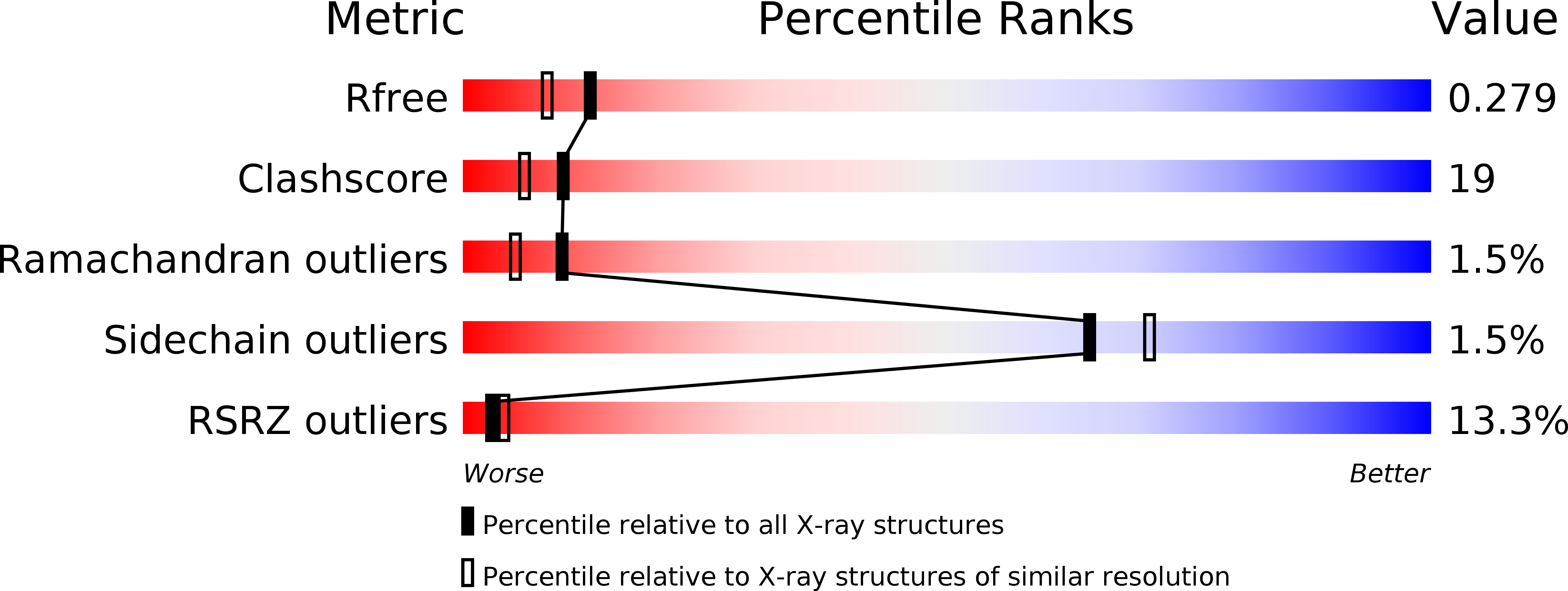
Resolution:
2.10 Å
R-Value Free:
0.27
R-Value Work:
0.23
Space Group:
P 41 21 2
Macromolecular Entities
Polymer Type:polypeptide(L)
Molecule:EPOXIDE HYDROLASE EPHB
Gene (Uniprot):MT1988
Chain IDs:A
Chain Length:356
Number of Molecules:1
Biological Source:Mycobacterium tuberculosis
Ligand Molecules
Primary Citation
The molecular structure of epoxide hydrolase B from Mycobacterium tuberculosis and its complex with a urea-based inhibitor.
J.Mol.Biol. 381 897 912 (2008)
PMID: 18585390 DOI: 10.1016/j.jmb.2008.06.030

Abstact

Mycobacterium tuberculosis (Mtb), the intracellular pathogen that infects macrophages primarily, is the causative agent of the infectious disease tuberculosis in humans. The Mtb genome encodes at least six epoxide hydrolases (EHs A to F). EHs convert epoxides to trans-dihydrodiols and have roles in drug metabolism as well as in the processing of signaling molecules. Herein, we report the crystal structures of unbound Mtb EHB and Mtb EHB bound to a potent, low-nanomolar (IC(50) approximately 19 nM) urea-based inhibitor at 2.1 and 2.4 A resolution, respectively. The enzyme is a homodimer; each monomer adopts the classical alpha/beta hydrolase fold that composes the catalytic domain; there is a cap domain that regulates access to the active site. The catalytic triad, comprising Asp104, His333 and Asp302, protrudes from the catalytic domain into the substrate binding cavity between the two domains. The urea portion of the inhibitor is bound in the catalytic cavity, mimicking, in part, the substrate binding; the two urea nitrogen atoms donate hydrogen bonds to the nucleophilic carboxylate of Asp104, and the carbonyl oxygen of the urea moiety receives hydrogen bonds from the phenolic oxygen atoms of Tyr164 and Tyr272. The phenolic oxygen groups of these two residues provide electrophilic assistance during the epoxide hydrolytic cleavage. Upon inhibitor binding, the binding-site residues undergo subtle structural rearrangement. In particular, the side chain of Ile137 exhibits a rotation of around 120 degrees about its C(alpha)-C(beta) bond in order to accommodate the inhibitor. These findings have not only shed light on the enzyme mechanism but also have opened a path for the development of potent inhibitors with good pharmacokinetic profiles against all Mtb EHs of the alpha/beta type.

Legend

Protein

Chemical

Disease

Primary Citation of related structures