Deposition Date
2006-11-18
Release Date
2007-10-23
Last Version Date
2023-10-25
Entry Detail
PDB ID:
2E2Y
Keywords:
Title:
Crystal Structure of F43W/H64D/V68I Myoglobin
Biological Source:
Source Organism(s):
Physeter catodon (Taxon ID: 9755)
Expression System(s):
Method Details:
Experimental Method:
Resolution:
1.60 Å
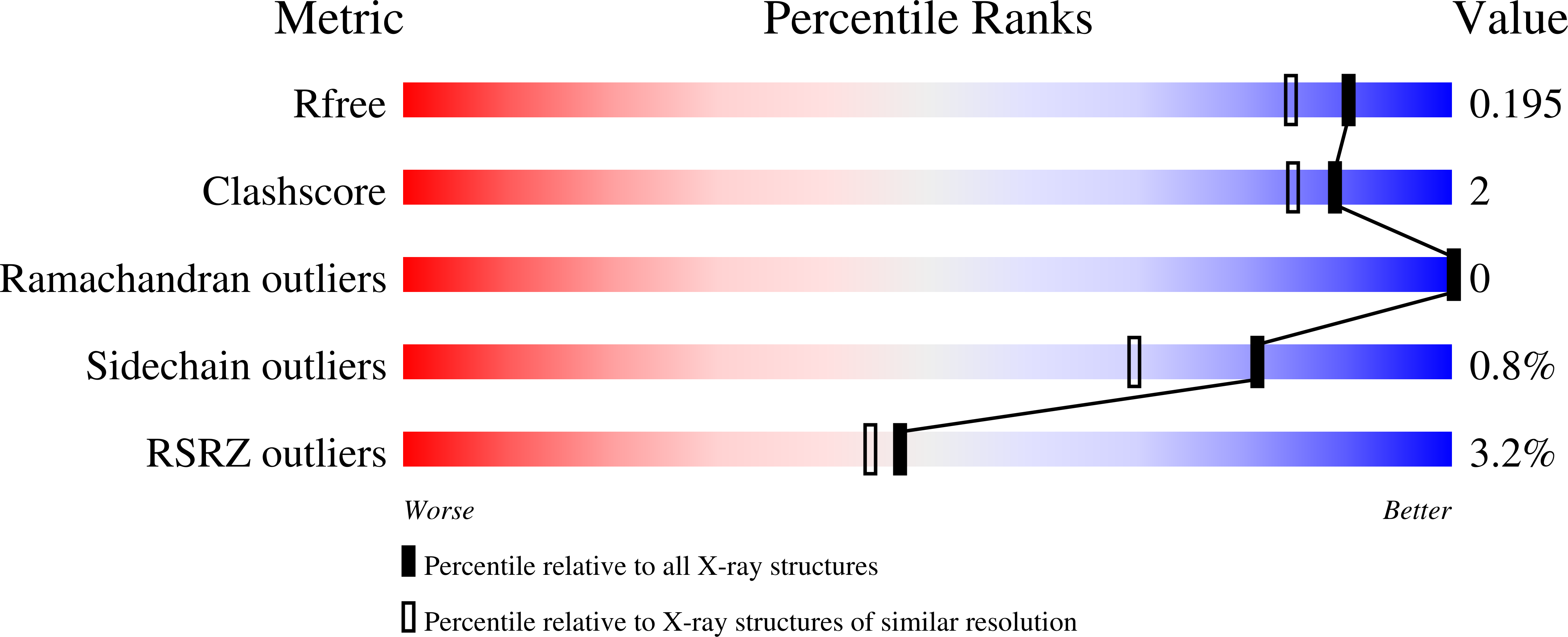
R-Value Free:
0.19
R-Value Work:
0.16
R-Value Observed:
0.16
Space Group:
P 6