2E26 image
Deposition Date 2006-11-08
Release Date 2007-05-22
Last Version Date 2024-11-06
Entry Detail
PDB ID:
2E26
Title:
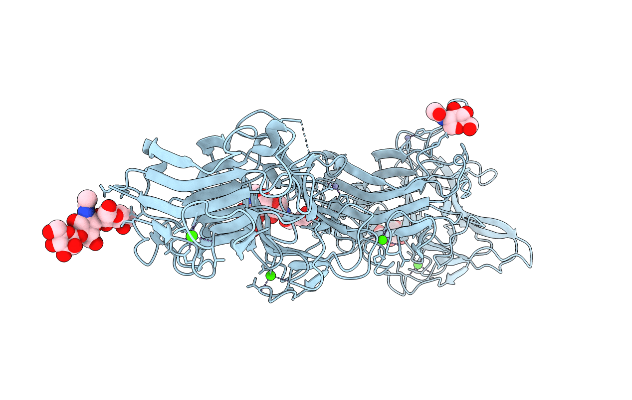
Crystal structure of two repeat fragment of reelin
Biological Source:
Source Organism:
Mus musculus (Taxon ID: 10090)
Host Organism:
Method Details:
Experimental Method:
Resolution:
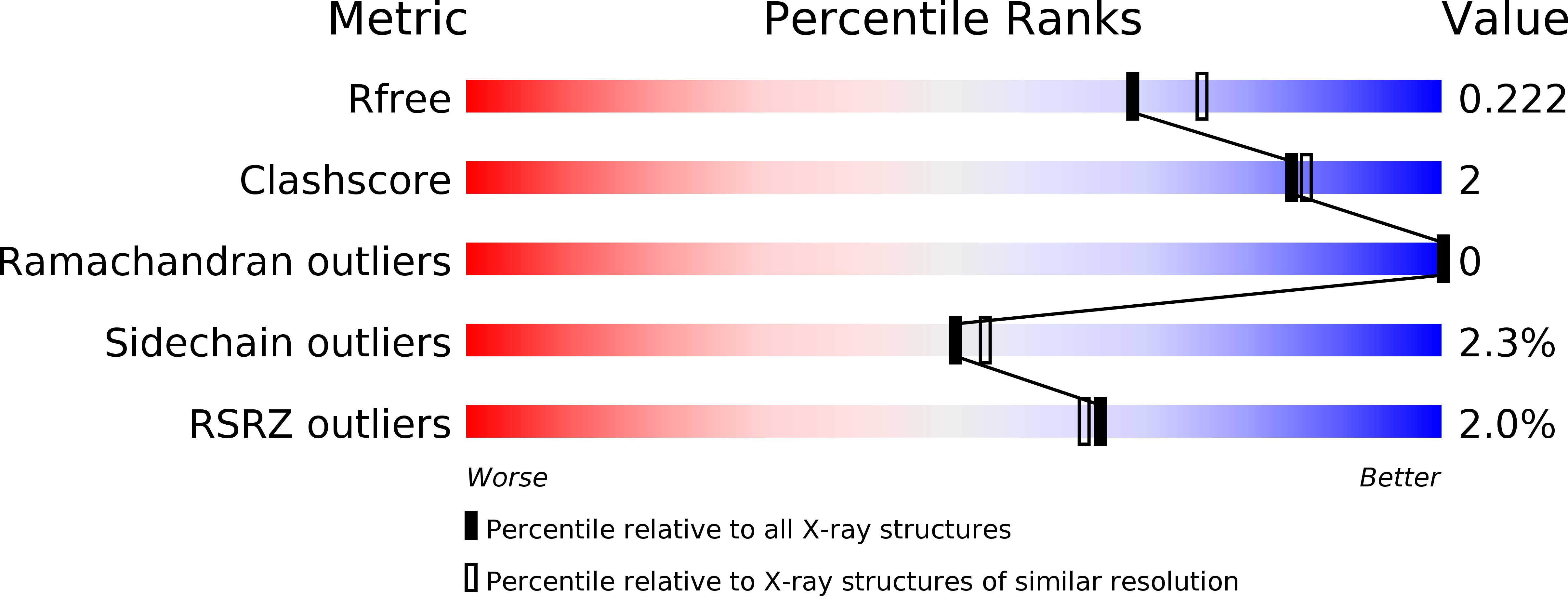
2.00 Å
R-Value Free:
0.21
R-Value Work:
0.17
R-Value Observed:
0.18
Space Group:
P 1 21 1
Macromolecular Entities
Structures with similar UniProt ID
Protein Blast
Polymer Type:polypeptide(L)
Molecule:Reelin
Gene (Uniprot):Reln
Chain IDs:A
Chain Length:725
Number of Molecules:1
Biological Source:Mus musculus
Modified Residue
Compound ID Chain ID Parent Comp ID Details 2D Image
ASN A ASN GLYCOSYLATION SITE
Primary Citation
Structure of a receptor-binding fragment of reelin and mutational analysis reveal a recognition mechanism similar to endocytic receptors.
Proc.Natl.Acad.Sci.Usa 104 9988 9993 (2007)
PMID: 17548821 DOI: 10.1073/pnas.0700438104

Abstact

Reelin, a large secreted protein implicated in the cortical development of the mammalian brain, is composed of eight tandem concatenations of "reelin repeats" and binds to neuronal receptors belonging to the low-density lipoprotein receptor gene family. We found that both receptor-binding and subsequent Dab1 phosphorylation occur solely in the segment spanning the fifth and sixth reelin repeats (R5-6). Monomeric fragment exhibited a suboptimal level of signaling activity and artificial oligomerization resulted in a 10-fold increase in activity, indicating the critical importance of higher-order multimerization in physiological reelin. A 2.0-A crystal structure from the R5-6 fragment revealed not only a unique domain arrangement wherein two repeats were aligned side by side with the same orientation, but also the unexpected presence of bound Zn ions. Structure-guided alanine mutagenesis of R5-6 revealed that two Lys residues (Lys-2360 and Lys-2467) constitute a central binding site for the low-density lipoprotein receptor class A module in the receptor, indicating a strong similarity to the ligand recognition mode shared among the endocytic lipoprotein receptors.

Legend

Protein

Chemical

Disease

Primary Citation of related structures
Feedback Form
Name
Email
Institute
Feedback