2E0T image
Deposition Date 2006-10-13
Release Date 2007-10-16
Last Version Date 2024-03-13
Entry Detail
PDB ID:
2E0T
Keywords:
Title:
Crystal structure of catalytic domain of dual specificity phosphatase 26, MS0830 from Homo sapiens
Biological Source:
Source Organism:
Homo sapiens (Taxon ID: 9606)
Method Details:
Experimental Method:
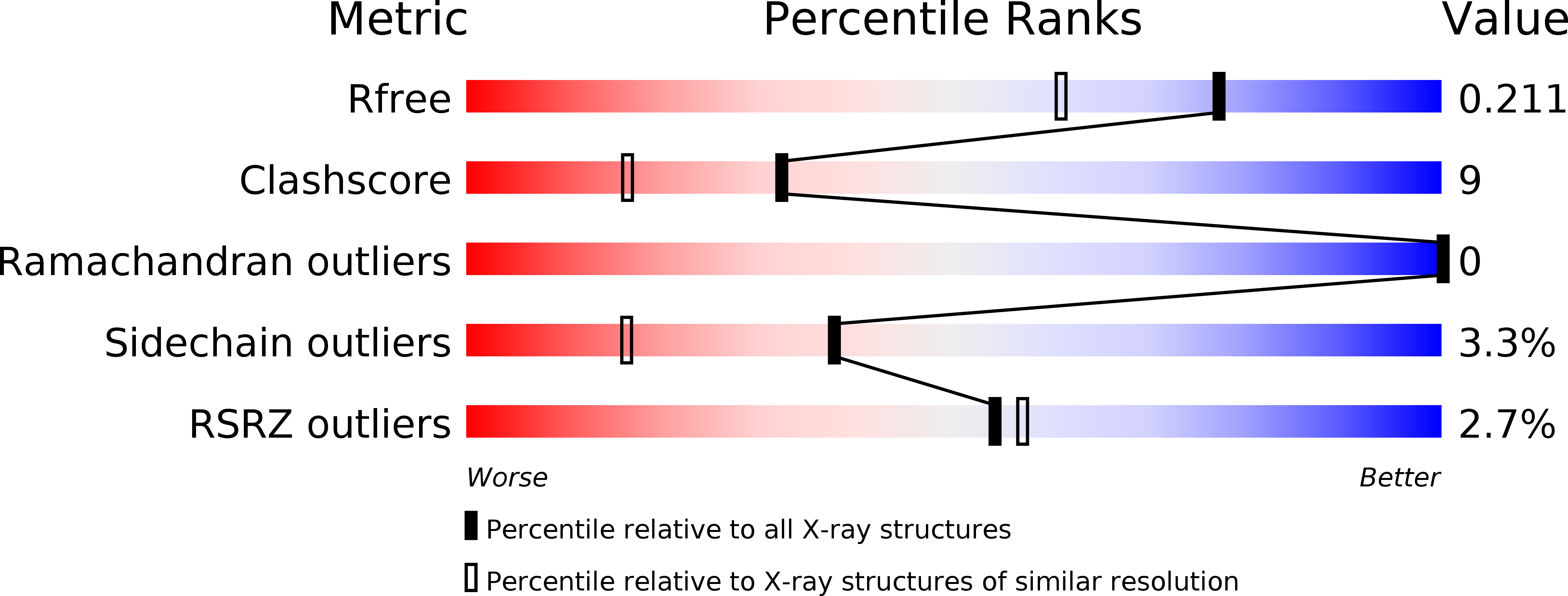
Resolution:
1.67 Å
R-Value Free:
0.21
R-Value Work:
0.17
R-Value Observed:
0.17
Space Group:
C 1 2 1
Macromolecular Entities
Structures with similar UniProt ID
Protein Blast
Polymer Type:polypeptide(L)
Molecule:Dual specificity phosphatase 26
Gene (Uniprot):DUSP26
Chain IDs:A
Chain Length:151
Number of Molecules:1
Biological Source:Homo sapiens
Primary Citation
High-resolution crystal structure of the catalytic domain of human dual-specificity phosphatase 26.
Acta Crystallogr.,Sect.D 69 1160 1170 (2013)
PMID: 23695260 DOI: 10.1107/S0907444913004770

Abstact

Dual-specificity phosphatases (DUSPs) play an important role in regulating cellular signalling pathways governing cell growth, differentiation and apoptosis. Human DUSP26 inhibits the apoptosis of cancer cells by dephosphorylating substrates such as p38 and p53. High-resolution crystal structures of the DUSP26 catalytic domain (DUSP26-C) and its C152S mutant [DUSP26-C (C152S)] have been determined at 1.67 and 2.20 Å resolution, respectively. The structure of DUSP26-C showed a novel type of domain-swapped dimer formed by extensive crossover of the C-terminal α7 helix. Taken together with the results of a phosphatase-activity assay, structural comparison with other DUSPs revealed that DUSP26-C adopts a catalytically inactive conformation of the protein tyrosine phosphate-binding loop which significantly deviates from that of canonical DUSP structures. In particular, a noticeable difference exists between DUSP26-C and the active forms of other DUSPs at the hinge region of a swapped C-terminal domain. Additionally, two significant gaps were identified between the catalytic core and its surrounding loops in DUSP26-C, which can be exploited as additional binding sites for allosteric enzyme regulation. The high-resolution structure of DUSP26-C may thus provide structural insights into the rational design of DUSP26-targeted anticancer drugs.

Legend

Protein

Chemical

Disease

Primary Citation of related structures