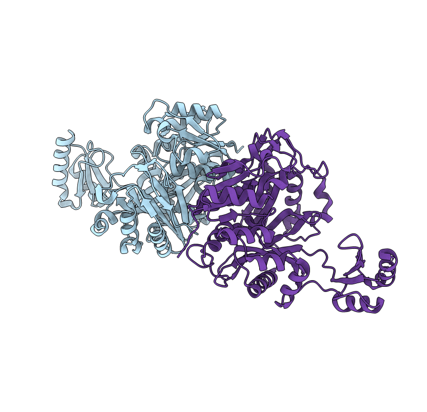
2DZD image
Entry Detail
PDB ID:
2DZD
Keywords:
Title:
Crystal structure of the biotin carboxylase domain of pyruvate carboxylase
Biological Source:
Host Organism:
PDB Version:
Deposition Date:
2006-09-27
Release Date:
2007-09-25
Method Details:
Experimental Method:
Resolution:
2.40 Å
R-Value Free:
0.29
R-Value Work:
0.23
Space Group:
P 21 21 21
Macromolecular Entities
Polymer Type:polypeptide(L)
Description:pyruvate carboxylase
Chain IDs:A, B
Chain Length:461
Number of Molecules:2
Biological Source:Geobacillus thermodenitrificans
Primary Citation
Structure of the biotin carboxylase domain of pyruvate carboxylase from Bacillus thermodenitrificans
ACTA CRYSTALLOGR.,SECT.D 63 885 890 (2007)
PMID: 17642515 DOI: 10.1107/S0907444907029423

Abstact

The biotin carboxylase (BC) domain of pyruvate carboxylase (PC) from Bacillus thermodenitrificans (BC-bPC) was crystallized in an orthorhombic form (space group P2(1)2(1)2(1)), with unit-cell parameters a = 79.6, b = 116.0, c = 115.7 A. Two BC protomers are contained in the asymmetric unit. Diffraction data were collected at 100 K and the crystal structure was solved by the molecular-replacement method and refined against reflections in the 20.0-2.4 A resolution range, giving an R factor of 0.235 and a free R factor of 0.292. The overall structure of BC-bPC is similar to those of the BC subunits of Aquifex aeolicus PC (BC-aPC) and Escherichia coli ACC (BC-eACC). The crystal structure revealed that BC-bPC forms a unique dimeric quaternary structure, which might be caused as a result of the division of the BC domain from the rest of the protein. The position of domain B in BC-bPC differs from those in other enzymes of similar structure (BC-aPC and BC-eACC).

Legend

Protein

Chemical

Disease

Primary Citation of related structures