Deposition Date
2006-08-17
Release Date
2007-02-17
Last Version Date
2024-05-29
Entry Detail
PDB ID:
2DWV
Keywords:
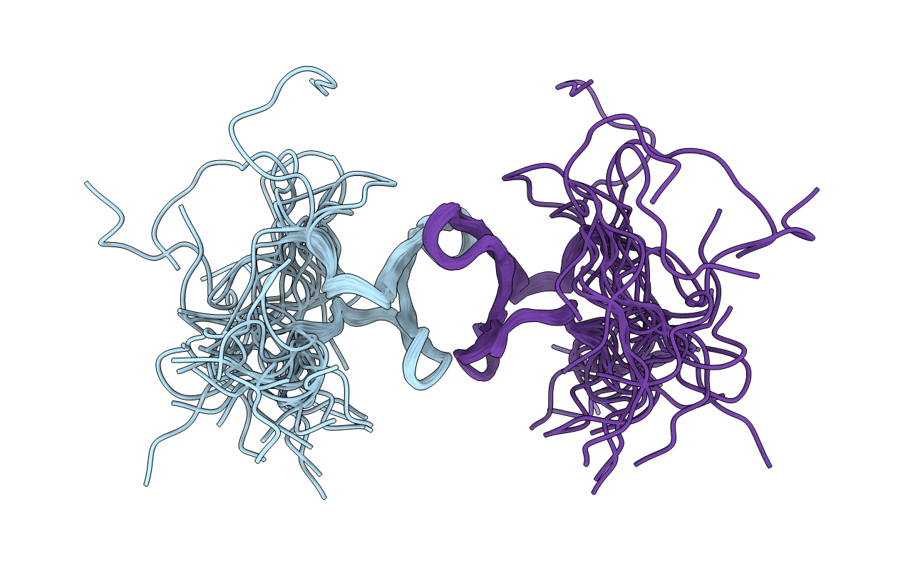
Title:
Solution structure of the second WW domain from mouse salvador homolog 1 protein (mWW45)
Biological Source:
Source Organism(s):
Mus musculus (Taxon ID: 10090)
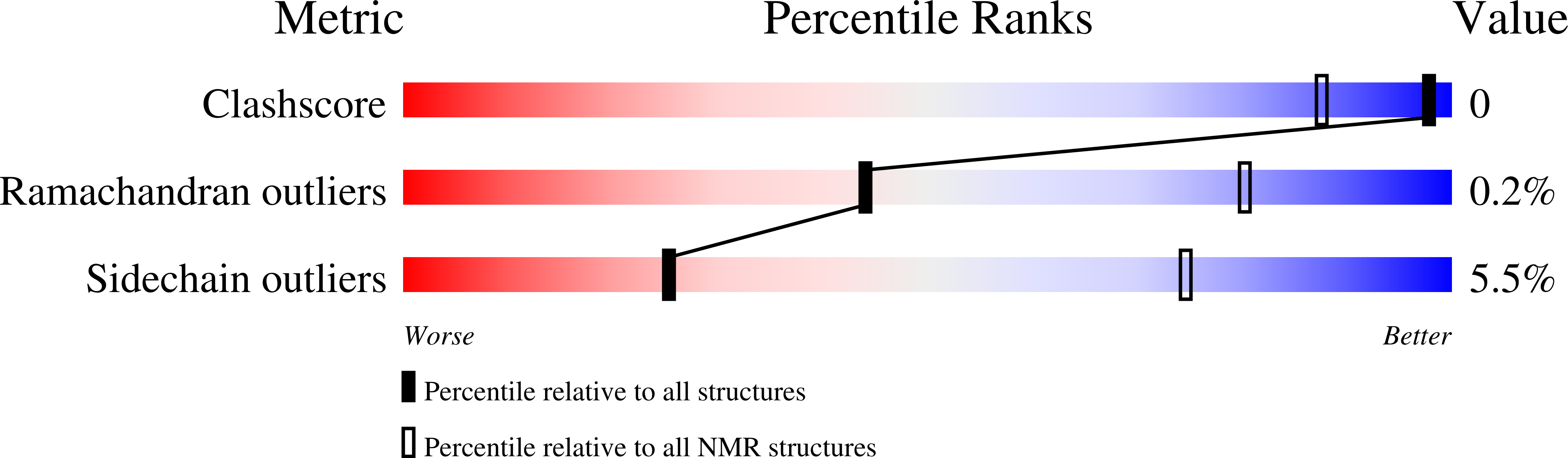
Method Details:
Experimental Method:
Conformers Calculated:
100
Conformers Submitted:
20
Selection Criteria:
structures with the least restraint violations