Deposition Date
2006-08-02
Release Date
2007-06-26
Last Version Date
2024-05-29
Entry Detail
PDB ID:
2DW3
Keywords:
Title:
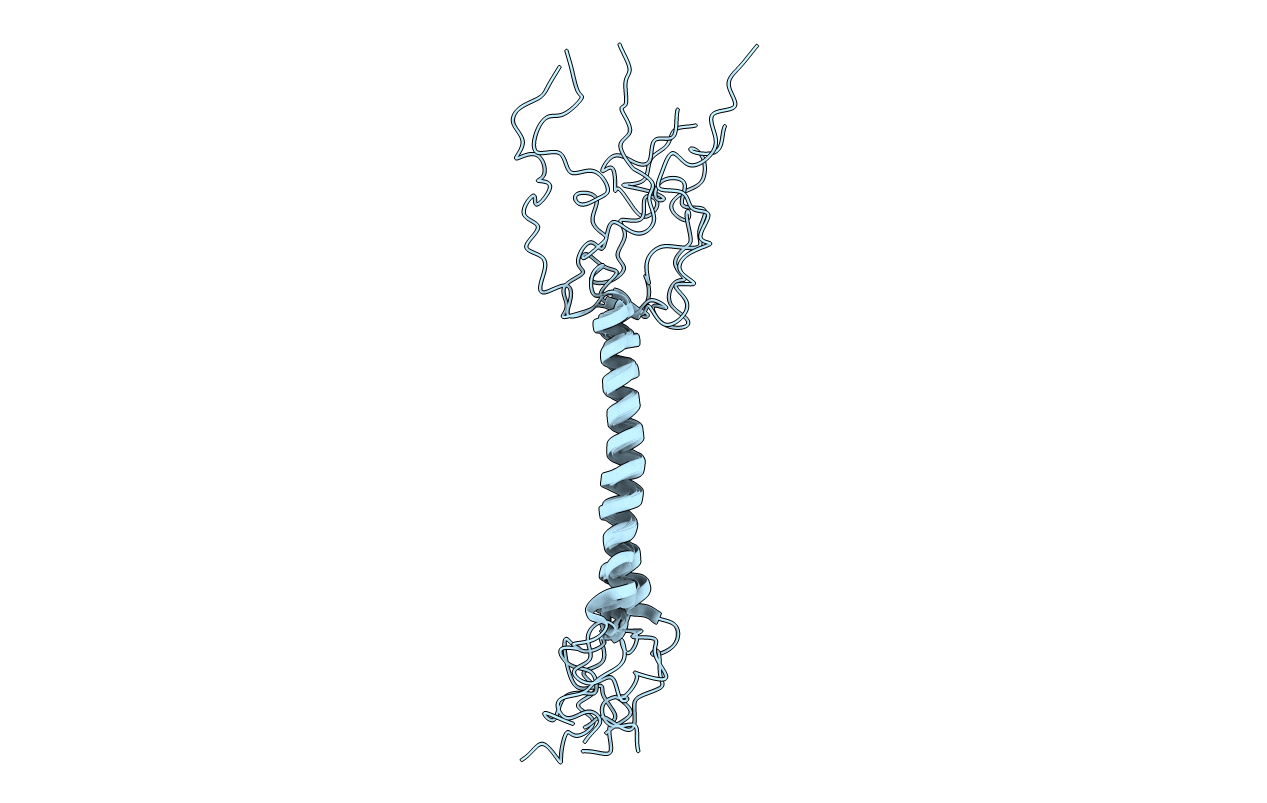
Solution structure of the Rhodobacter sphaeroides PufX membrane protein
Biological Source:
Source Organism(s):
Rhodobacter sphaeroides (Taxon ID: 1063)
Expression System(s):
Method Details:
Experimental Method:
Conformers Calculated:
200
Conformers Submitted:
7
Selection Criteria:
structures with the lowest energy