Deposition Date
2006-07-31
Release Date
2006-11-07
Last Version Date
2023-10-25
Entry Detail
PDB ID:
2DVF
Keywords:
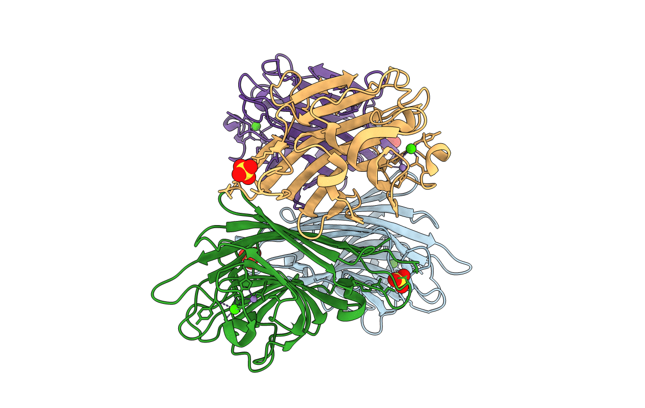
Title:
Crystals of peanut lectin grown in the presence of GAL-ALPHA-1,3-GAL-BETA-1,4-GAL
Biological Source:
Source Organism(s):
Arachis hypogaea (Taxon ID: 3818)
Method Details:
Experimental Method:
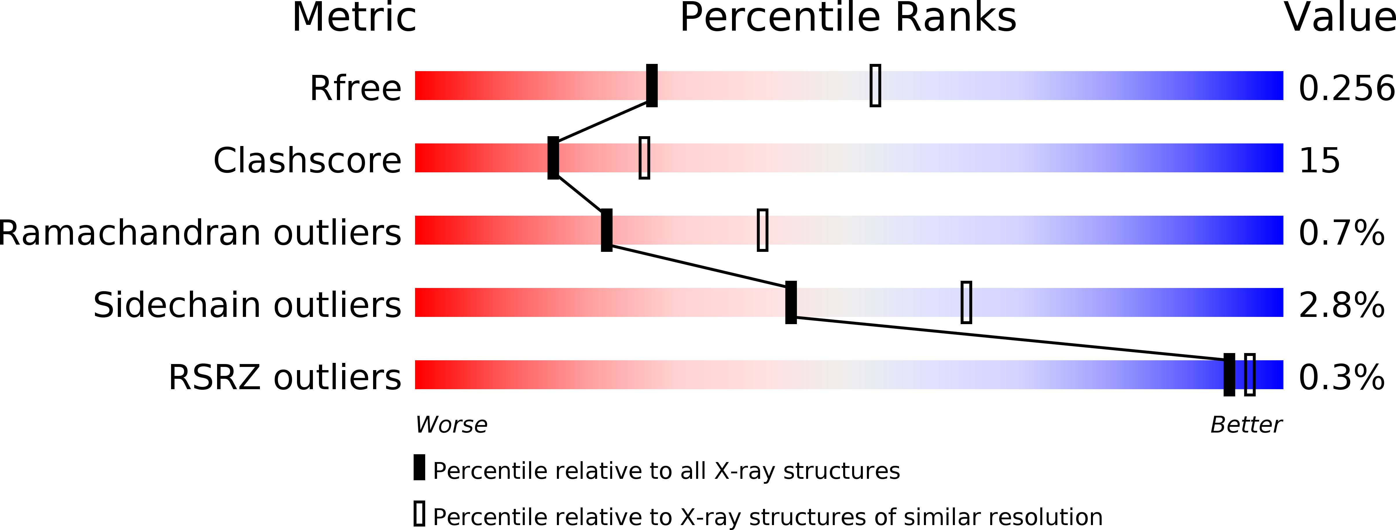
Resolution:
2.74 Å
R-Value Free:
0.26
R-Value Work:
0.2
R-Value Observed:
0.2
Space Group:
P 21 21 2