2DUP image
Deposition Date 2006-07-25
Release Date 2007-07-24
Last Version Date 2024-11-20
Entry Detail
PDB ID:
2DUP
Title:
Crystal structure of VIP36 exoplasmic/lumenal domain, metal-free form
Biological Source:
Source Organism:
Method Details:
Experimental Method:
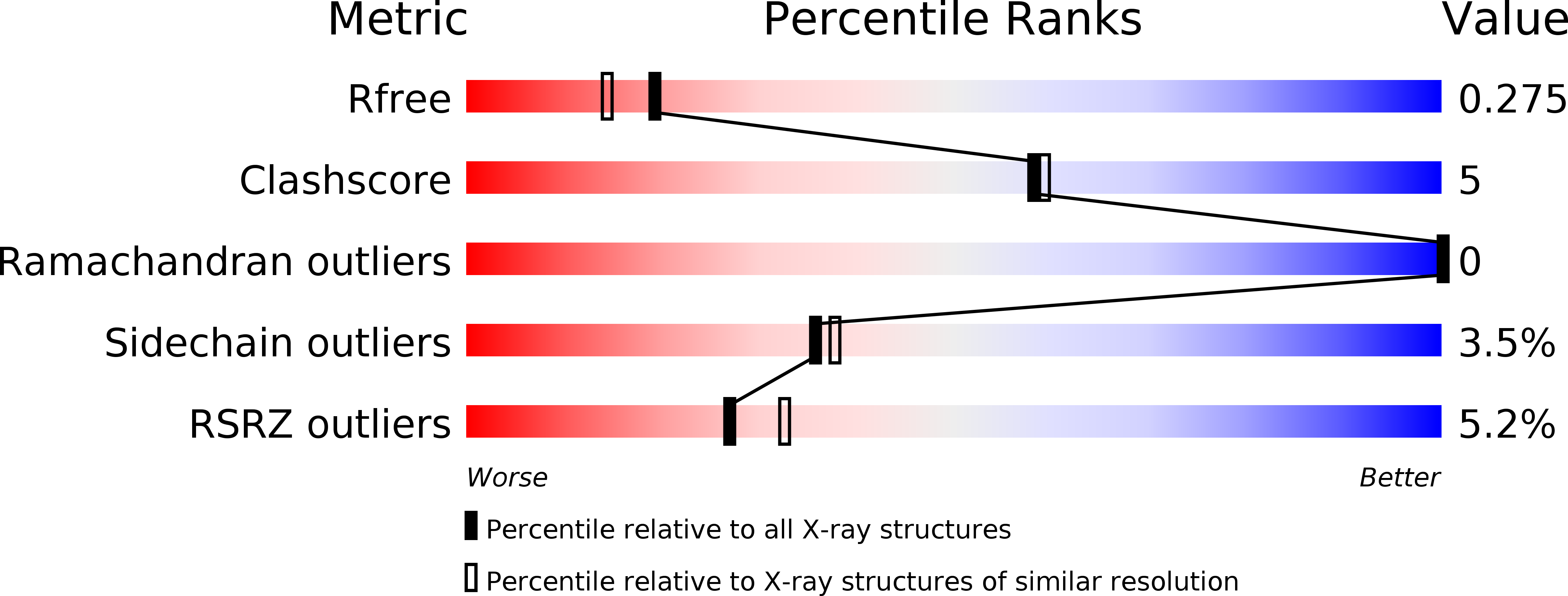
Resolution:
2.10 Å
R-Value Free:
0.27
R-Value Work:
0.22
R-Value Observed:
0.22
Space Group:
C 1 2 1
Macromolecular Entities
Polymer Type:polypeptide(L)
Molecule:Vesicular integral-membrane protein VIP36
Gene (Uniprot):LMAN2
Chain IDs:A, B
Chain Length:253
Number of Molecules:2
Biological Source:Canis lupus familiaris
Primary Citation
Structural basis for recognition of high mannose type glycoproteins by mammalian transport lectin VIP36
J.Biol.Chem. 282 28246 28255 (2007)
PMID: 17652092 DOI: 10.1074/jbc.M703064200

Abstact

VIP36 functions as a transport lectin for trafficking certain high mannose type glycoproteins in the secretory pathway. Here we report the crystal structure of VIP36 exoplasmic/luminal domain comprising a carbohydrate recognition domain and a stalk domain. The structures of VIP36 in complex with Ca(2+) and mannosyl ligands are also described. The carbohydrate recognition domain is composed of a 17-stranded antiparallel beta-sandwich and binds one Ca(2+) adjoining the carbohydrate-binding site. The structure reveals that a coordinated Ca(2+) ion orients the side chains of Asp(131), Asn(166), and His(190) for carbohydrate binding. This result explains the Ca(2+)-dependent carbohydrate binding of this protein. The Man-alpha-1,2-Man-alpha-1,2-Man, which corresponds to the D1 arm of high mannose type glycan, is recognized by eight residues through extensive hydrogen bonds. The complex structures reveal the structural basis for high mannose type glycoprotein recognition by VIP36 in a Ca(2+)-dependent and D1 arm-specific manner.

Legend

Protein

Chemical

Disease

Primary Citation of related structures