Deposition Date
2006-07-23
Release Date
2007-07-10
Last Version Date
2024-11-13
Entry Detail
PDB ID:
2DUF
Keywords:
Title:
crystal structure of a green fluorescent protein variant S65T/H148D at pH 5.6
Biological Source:
Source Organism:
Aequorea victoria (Taxon ID: 6100)
Host Organism:
Method Details:
Experimental Method:
Resolution:
1.50 Å
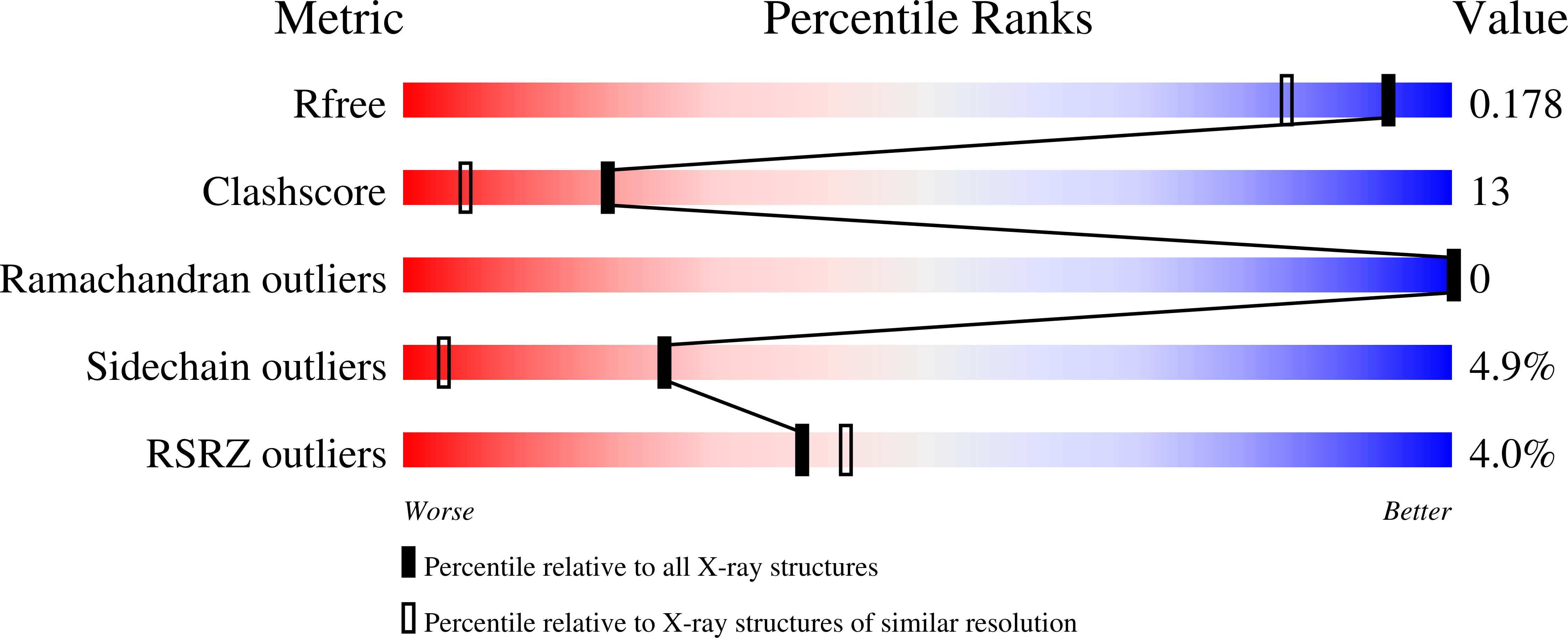
R-Value Free:
0.24
R-Value Work:
0.17
R-Value Observed:
0.17
Space Group:
P 21 21 21