2DUB image
Deposition Date 1997-04-28
Release Date 1998-04-29
Last Version Date 2024-12-25
Entry Detail
PDB ID:
2DUB
Keywords:
Title:
ENOYL-COA HYDRATASE COMPLEXED WITH OCTANOYL-COA
Biological Source:
Source Organism:
Method Details:
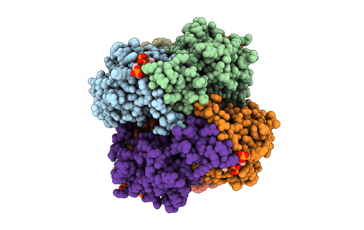
Experimental Method:
Resolution:
2.40 Å
R-Value Free:
0.26
R-Value Work:
0.20
Space Group:
P 21 21 21
Macromolecular Entities
Polymer Type:polypeptide(L)
Molecule:2-ENOYL-COA HYDRATASE
Gene (Uniprot):Echs1
Chain IDs:A, B, C, D, E, F
Chain Length:261
Number of Molecules:6
Biological Source:Rattus norvegicus
Ligand Molecules
Primary Citation
The crystal structure of enoyl-CoA hydratase complexed with octanoyl-CoA reveals the structural adaptations required for binding of a long chain fatty acid-CoA molecule.
J.Mol.Biol. 275 847 859 (1998)
PMID: 9480773 DOI: 10.1006/jmbi.1997.1491

Abstact

The structure of the hexameric rat mitochondrial enoyl-Coenzyme A (CoA) hydratase, co-crystallised with the inhibitor octanoyl-CoA, has been refined at a resolution of 2.4 A. Enoyl-CoA hydratase catalyses the hydration of 2,3-unsaturated enoyl-CoA thioesters. In the crystal structure only four of the six active sites of the hexamer in the asymmetric unit are occupied with a ligand molecule, showing an unliganded and a liganded active site within the same crystal form. While the protein assembly and fold is identical to the previously solved acetoacetyl-CoA complex, differences are observed close to the fatty acid binding pocket due to the different nature of the ligands. The fatty acid tail of octanoyl-CoA is bound in an extended conformation. This is possible because a high B-factor loop, which separates in the acetoacetyl-CoA complex the binding pocket of the acetoacetyl-CoA fatty acid tail from the intertrimer space, has moved aside to allow binding of the longer octanoyl-CoA moiety. The movement of this loop opens a tunnel which traverses the complete subunit from the solvent space to the intertrimer space. The conformation of the catalytic residues is identical, in both structures as well as in the liganded and the unliganded active sites. In the unliganded active sites a water molecules is bound between the two catalytic glutamate, residues Glu144 and Glu164. After superposition of a liganded active site on an unliganded active site this water molecule is close to the carbon centre that becomes hydroxylated in the hydratase reaction. These findings support the idea that the active site is rigid and that the catalytic residues and the water molecule, as seen in the unliganded active site, are pre-positioned for very efficient catalysis.

Legend

Protein

Chemical

Disease

Primary Citation of related structures